You have no items in your shopping cart.
SEMA3A antibody
SKU: orb11359
Featured
Description
Images & Validation
−Item 1 of 15
| Tested Applications | ICC, IF, IHC-P, WB |
|---|---|
| Dilution range | WB: 1:100-800, IHC-P: 1: 100-500 |
| Reactivity | Human, Mouse, Rat |
Key Properties
−| Host | Rabbit |
|---|---|
| Clonality | Polyclonal |
| Isotype | IgG |
| Immunogen | KLH conjugated synthetic peptide derived from human SEMA3A. Please contact us for the exact immunogen sequence. The peptide is available as orb374747. |
| Target | SEMA3A |
| Molecular Weight | 89 kDa |
| Purity | Polyclonal antibodies are purified by peptide affinity chromatography |
Storage & Handling
−| Storage | Maintain refrigerated at 2-8°C for up to 2 weeks. For long term storage store at -20°C in small aliquots to prevent freeze-thaw cycles. |
|---|---|
| Form/Appearance | 10 mM PBS, 0.02% sodium azide |
| Concentration | - 100 μg (in 200 μl): 0.5 mg/ml- 200 μg (in 400 μl): 0.5 mg/ml |
| Disclaimer | For research use only |
Alternative Names
−anti Coll 1 antibody, anti Coll1 antibody, anti HH16 antibody, anti Hsema I antibody, anti Hsema III antibody, anti Sema III antibody, anti SEMA1 antibody, anti SEMA1-PEN antibody, anti SEMA3A antibody, anti SEMAD antibody, anti SEMAIII antibody, anti SEMAL antibody, anti Semaphorin 3A precursor antibody, anti Semaphorin III antibody, anti Semaphorin-3A precursor antibody, anti Semaphorin3A antibody, anti SemD antibody
Similar Products
−- Item 1 of 1
- Item 1 of 1
- Item 1 of 1
- Item 1 of 3
SEMA3A Antibody [orb415845]
ELISA, IHC, WB
Human, Mouse, Rat
Rabbit
Polyclonal
Unconjugated
100 μg, 50 μg - Item 1 of 2
SEMA3A Antibody (Center) [orb1928916]
IHC-P, WB
Gallus, Mouse, Rat
Human
Rabbit
Polyclonal
Unconjugated
100 μl, 50 μl


Quality Guarantee
Explore bioreagents carefree to elevate your research. All our products are rigorously tested for performance. If a product does not perform as described on its datasheet, our scientific support team will provide expert troubleshooting, a prompt replacement, or a refund. For full details, please see our Terms & Conditions and Buying Guide. Contact us at [email protected].

Immunohistochemical staining of paraffin embedded human breast cancer tissue using Semaphorin 3A antibody (primary antibody at 1:200)

IHC-P image of mouse lung tissue using anti-Semaphorin 3A (dilution of primary antibody at 1:100)
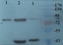
WB analysis of Mouse lung (Lane 1), Mouse liver (Lane 2), Mouse kidney (Lane 3) using Semaphorin 3A antibody (primary antibody at 2 ug/ml)

Immunofluorescence analysis of mouse lung tissue using Semaphorin 3A antibody (dilution of primary antibody - 1:200)

Immunohistochemical staining of mouse lung tissue using Semaphorin 3A antibody (dilution of primary antibody - 1:200)
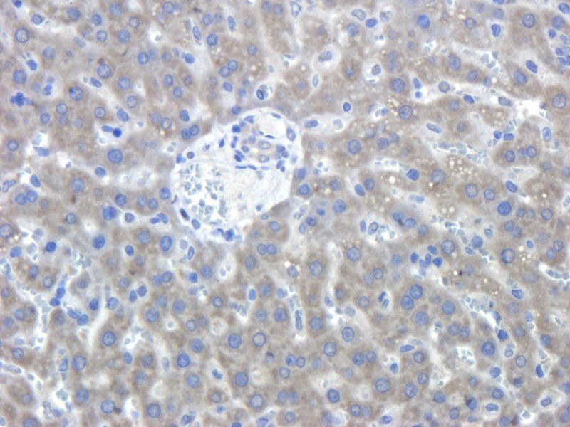
IHC-P staining of rat liver tissue using anti-Semaphorin 3A (dilution at 1:100)

Immunohistochemical staining of paraffin embedded rat liver tissue using anti-Semaphorin 3A (primary antibody at 1:200)
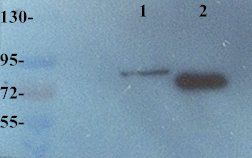
Western blot analysis of Mouse liver (Lane 1), Mouse lung (Lane 2) using anti Semaphorin 3A (dilution of primary antibody at 2 ug/ml)
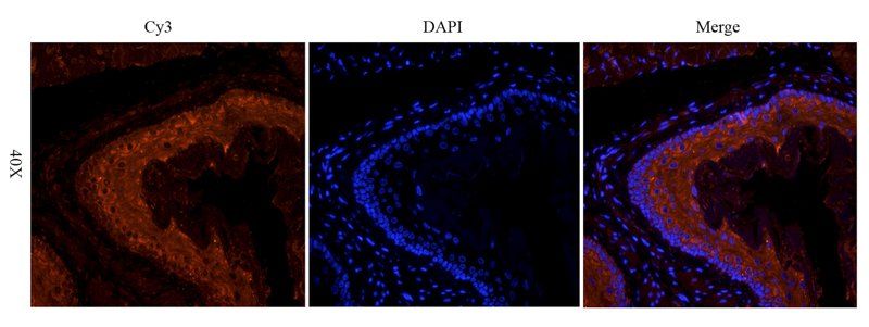
Immunofluorescence image of mouse lung tissue using anti-Semaphorin 3A (dilution at 1:200)
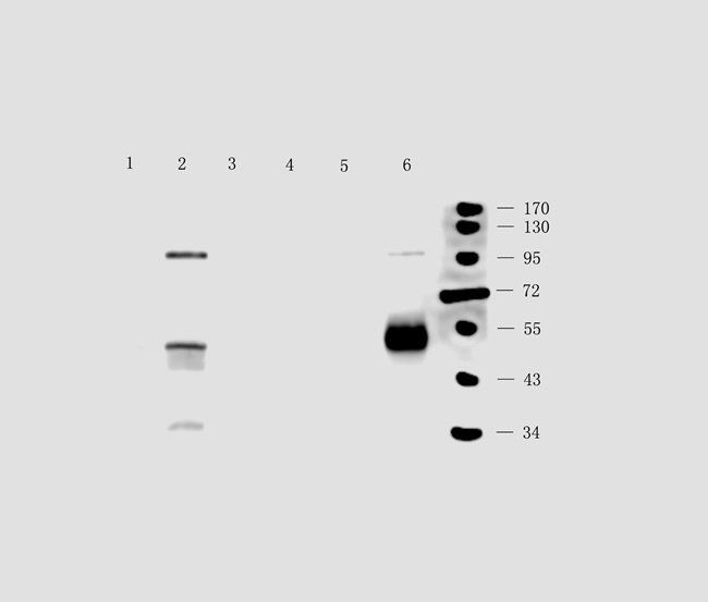
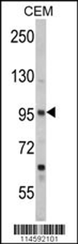
WB analysis of rat lung (lane 1), rat brain (lane 2), mouse brain (lane3),rat kidney (lane4), rat skin (lane 5), human breast cancer (lane 6) tissue using anti-SEM3A (0.5 ug/ml)

IHC-P staining of mouse stomach tissue using SEM3A antibody (2.5 ug/ml)
IHC-P image of mouse skin tissue using anti-SEM3A (2.5 ug/ml)

Immunohistochemical staining of mouse skin tissue using anti-SEM3A (2.5 ug/ml)
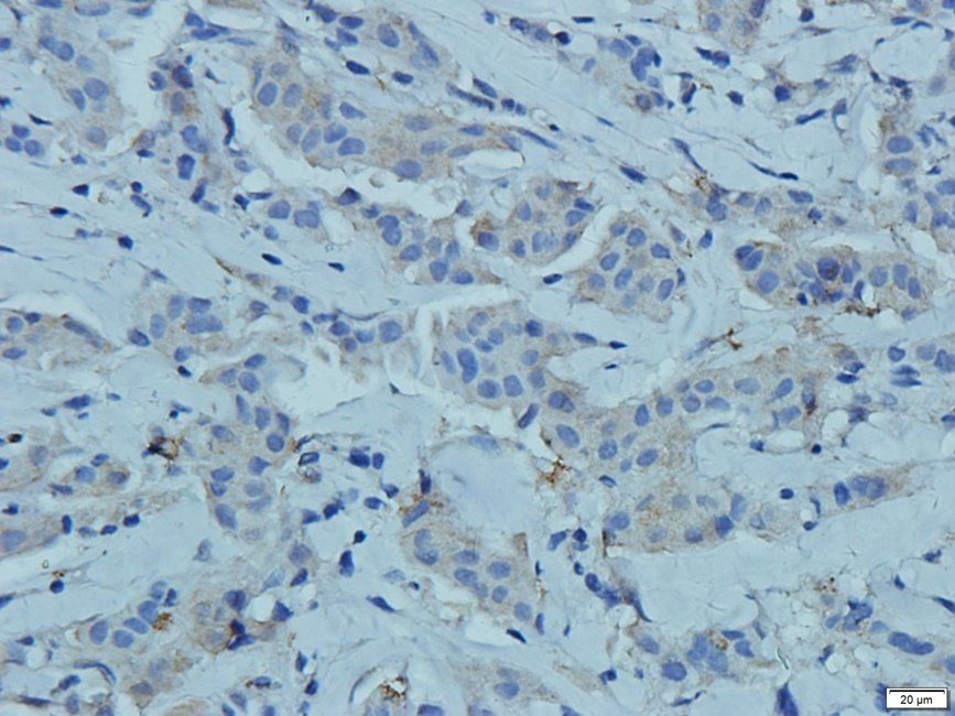
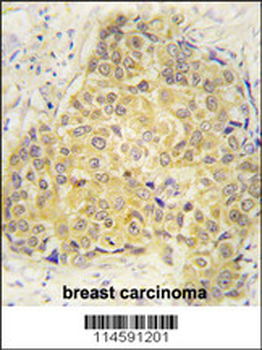
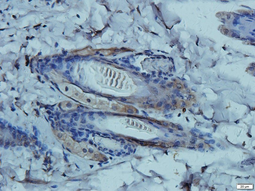
IHC-P staining of human breast cancer tissue using anti-SEM3A (2.5 ug/ml)
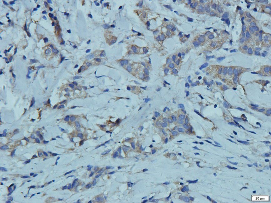
IHC-P image of human breast cancer tissue using anti-SEM3A (2.5 ug/ml)
Quick Database Links
Documents Download
Datasheet
Product Information
Request a Document
Protocol Information
WB
Western Blot (IB, immunoblot)
IHC-P
Immunohistochemistry Paraffin
IF
Immunofluorescence
ICC
Immunocytochemistry
SEMA3A antibody (orb11359)

- 0.0
Based on 0 reviews
Participating in our Biorbyt product reviews program enables you to support fellow scientists by sharing your firsthand experience with our products.
Login to Submit a ReviewAvailable Sizes
Select a size below