You have no items in your shopping cart.
Cart summary
Item 1 of 5
Item 1 of 5
WT1 Antibody (Center E361)
Catalog Number: orb1937831

| Catalog Number | orb1937831 |
|---|---|
| Category | Antibodies |
| Description | Affinity Purified Rabbit Polyclonal Antibody (Pab) |
| Species/Host | Rabbit |
| Clonality | Polyclonal |
| Clone Number | RB18841 |
| Tested applications | FC, IF, IHC-P, WB |
| Predicted Reactivity | Other, Porcine, Rat |
| Reactivity | Human, Mouse |
| Isotype | Rabbit IgG |
| Antibody Type | Primary Antibody |
| Dilution range | IF: 1:10~50, WB: 1:1000, WB: 1:1000, IHC-P: 1:50~100, FC: 1:25 |
| Form/Appearance | Purified polyclonal antibody supplied in PBS with 0.09% (W/V) sodium azide. This antibody is purified through a protein A column, followed by peptide affinity purification. |
| Conjugation | Unconjugated |
| MW | 49188 Da |
| Target | This WT1 antibody is generated from rabbits immunized with a KLH conjugated synthetic peptide between 346-375 amino acids from the Central region of human WT1. |
| UniProt ID | P19544 |
| NCBI | NP_077742.2, NP_001185481.1, NP_000369.3, NP_077744.3, NP_001185480.1 |
| Storage | Maintain refrigerated at 2-8°C for up to 2 weeks. For long term storage store at -20°C in small aliquots to prevent freeze-thaw cycles |
| Alternative names | Wilms tumor protein, WT33, WT1 Read more... |
| Note | For research use only |
| Expiration Date | 12 months from date of receipt. |

All lanes: Anti-WT1 Antibody (Center E361) at 1:1000 dilution. Lane 1: HT-1080 whole cell lysate. Lane 2: K562 whole cell lysate. Lane 3: MOLT-4 whole cell lysate. Lane 4: Mouse kidney lysate.Lysates/proteins at 20 µg per lane. Secondary Goat Anti-Rabbit IgG, (H+L), Peroxidase conjugated at 1/10000 dilution. Predicted band size: 49 kDa. Blocking/Dilution buffer: 5% NFDM/TBST.
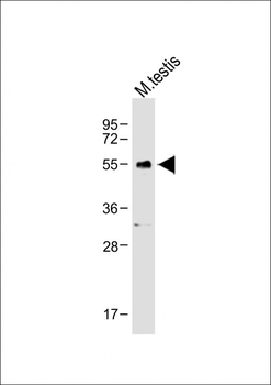
Anti-WT1 Antibody (Center E361) at 1:1000 dilution + Mouse testis lysate.Lysates/proteins at 20 µg per lane. Secondary Goat Anti-Rabbit IgG, (H+L), Peroxidase conjugated at 1/10000 dilution. Predicted band size: 49 kDa. Blocking/Dilution buffer: 5% NFDM/TBST.
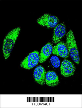
Confocal immunofluorescent analysis of WT1 Antibody (Center E361) with MCF-7 cell followed by Alexa Fluor 488-conjugated goat anti-rabbit lgG (green). DAPI was used to stain the cell nuclear (blue).
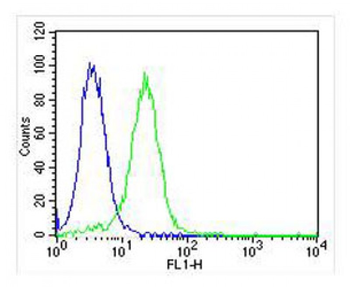
Overlay histogram showing Hela cells (green line). The cells were fixed with 2% paraformaldehyde (10 min) and then permeabilized with 90% methanol for 10 min. The cells were then icubated in 2% bovine serum albumin to block non-specific protein-protein interactions followed by the antibody (1:25 dilution) for 60 min at 37°C. The secondary antibody used was Goat-Anti-Rabbit IgG, DyLight 488 Conjugated Highly Cross-Adsorbed at 1/400 dilution for 40 min at 37°C. Isotype control antibody (blue line) was rabbit IgG1 (1 μg/1x10^6 cells) used under the same conditions. Acquisition of > 10000 events was performed.
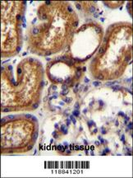
WT1 Antibody (Center E361) immunohistochemistry analysis in formalin fixed and paraffin embedded human kidney tissue followed by peroxidase conjugation of the secondary antibody and DAB staining. This data demonstrates the use of WT1 Antibody (Center E361) for immunohistochemistry. Clinical relevance has not been evaluated.

WT1 Antibody (Center E361) [orb1168020]
FC, IF, IHC-P, WB
Human
Rabbit
Polyclonal
Unconjugated
100 μl, 30 μlWT1 (Center E361) Antibody [orb2629917]
FC, ICC, IHC, WB
Human, Mouse, Rat
Rabbit
Polyclonal
Unconjugated
100 μl
