You have no items in your shopping cart.
Cart summary
Item 1 of 5
Item 1 of 5
Vinculin Antibody
Catalog Number: orb1925741

| Catalog Number | orb1925741 |
|---|---|
| Category | Antibodies |
| Description | Purified Rabbit Polyclonal Antibody (Pab) |
| Species/Host | Rabbit |
| Clonality | Polyclonal |
| Clone Number | RB55983 |
| Tested applications | FC, IHC-P, WB |
| Reactivity | Human, Mouse, Rat |
| Isotype | Rabbit IgG |
| Dilution range | WB: 1:2000, IHC-P: 1:25, IHC-P: 1:25, FC: 1:25, FC: 1:25 |
| Form/Appearance | Purified polyclonal antibody supplied in PBS with 0.09% (W/V) sodium azide. This antibody is purified through a protein A column, followed by peptide affinity purification. |
| Conjugation | Unconjugated |
| MW | 116717 Da |
| Target | This antibody is generated from a rabbit immunized with a KLH conjugated synthetic peptide between 903-937 amino acids from mouse. |
| UniProt ID | Q64727 |
| Storage | Maintain refrigerated at 2-8°C for up to 2 weeks. For long term storage store at -20°C in small aliquots to prevent freeze-thaw cycles |
| Alternative names | Vinculin, Metavinculin, Vcl Read more... |
| Note | For research use only |
| Expiration Date | 12 months from date of receipt. |

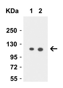
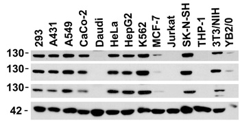
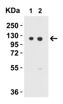
All lanes: Anti-Vinculin at 1:2000 dilution. Lane 1: human skeletal muscle lysate. Lane 2: A431 whole cell lysate. Lane 3: Hela whole cell lysate. Lane 4: mouse kidney lysate. Lane 5: PC-12 whole cell lysate. Lane 6: HepG2 whole cell lysate. Lysates/proteins at 20 µg per lane. Secondary Goat Anti-Rabbit IgG, (H+L), Peroxidase conjugated at 1/10000 dilution. Predicted band size: 117 kDa. Blocking/Dilution buffer: 5% NFDM/TBST.
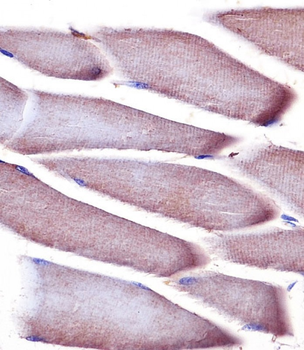
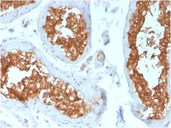
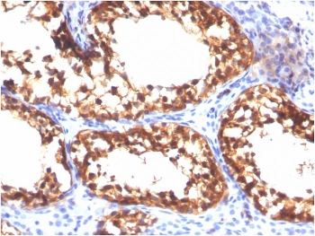
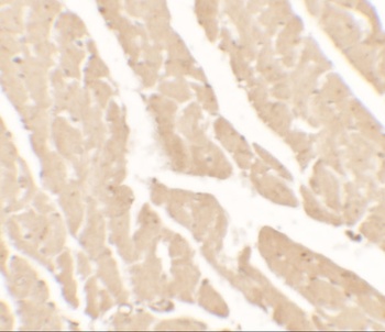
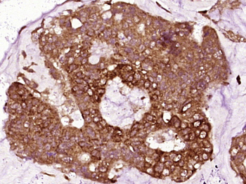
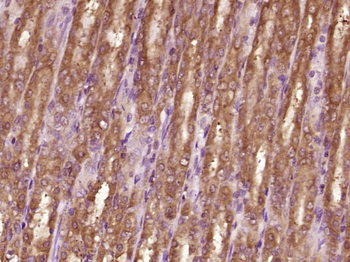
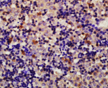
Staining Vinculin in mouse skeletal muscle tissue sections by Immunohistochemistry (IHC-P - paraformaldehyde-fixed, paraffin-embedded sections). Tissue was fixed with formaldehyde and blocked with 3% BSA for 0.5 hour at room temperature; antigen retrieval was by heat mediation with a citrate buffer (pH6). Samples were incubated with primary antibody (1/25) for 1 hours at 37°C. A undiluted biotinylated goat polyvalent antibody was used as the secondary antibody.
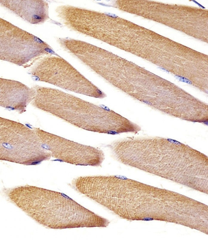
Staining Vinculin in mouse skeletal muscle tissue sections by Immunohistochemistry (IHC-P - paraformaldehyde-fixed, paraffin-embedded sections). Tissue was fixed with formaldehyde and blocked with 3% BSA for 0.5 hour at room temperature; antigen retrieval was by heat mediation with a citrate buffer (pH6). Samples were incubated with primary antibody (1/25) for 1 hours at 37°C. A undiluted biotinylated goat polyvalent antibody was used as the secondary antibody.
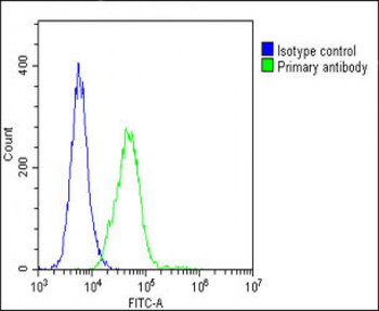
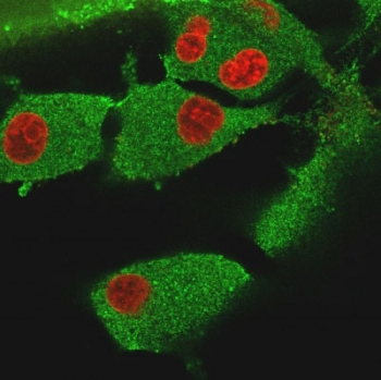
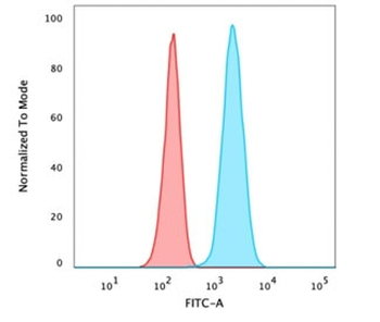
Overlay histogram showing C2C12 cells stained (green line). The cells were fixed with 2% paraformaldehyde (10 min) and then permeabilized with 90% methanol for 10 min. The cells were then icubated in 2% bovine serum albumin to block non-specific protein-protein interactions followed by the antibody (1:25 dilution) for 60 min at 37°C. The secondary antibody used was Goat-Anti-Rabbit IgG, DyLight 488 Conjugated Highly Cross-Adsorbed at 1/200 dilution for 40 min at 37°C. Isotype control antibody (blue line) was rabbit IgG1 (1 μg/1x10^6 cells) used under the same conditions. Acquisition of > 10000 events was performed.
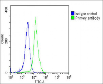
Overlay histogram showing NIH/3T3 cells stained (green line). The cells were fixed with 2% paraformaldehyde (10 min) and then permeabilized with 90% methanol for 10 min. The cells were then icubated in 2% bovine serum albumin to block non-specific protein-protein interactions followed by the antibody (1:25 dilution) for 60 min at 37°C. The secondary antibody used was Goat-Anti-Rabbit IgG, DyLight 488 Conjugated Highly Cross-Adsorbed at 1/200 dilution for 40 min at 37°C. Isotype control antibody (blue line) was rabbit IgG1 (1 μg/1x10^6 cells) used under the same conditions. Acquisition of > 10000 events was performed.
- Item 1 of 7
Vinculin Antibody [orb1240285]
ELISA, ICC, IF, WB
Human, Mouse, Rat
Rabbit
Polyclonal
Unconjugated
0.1 mg - Item 1 of 6
- Item 1 of 6
- Item 1 of 6
Vinculin Antibody [orb1240276]
ELISA, IF, IHC-P, WB
Gallus, Porcine
Human, Mouse, Rat
Rabbit
Polyclonal
Unconjugated
0.1 mg - Item 1 of 4
Vinculin Rabbit Polyclonal Antibody [orb100164]
FC, IF, IHC-Fr, IHC-P
Bovine, Canine, Equine, Gallus, Porcine, Rabbit
Human, Mouse, Rat
Rabbit
Polyclonal
Unconjugated
100 μl, 200 μl, 50 μl