You have no items in your shopping cart.
Cart summary
Item 1 of 3
Item 1 of 3
VAChT Antibody: Biotin
Catalog Number: orb149708

| Catalog Number | orb149708 |
|---|---|
| Category | Antibodies |
| Description | Mouse monoclonal to VAChT (Biotin). VAChT is a member of the vesicular amine transporter (VMAT) family. The encoded transmembrane protein transports acetylcholine into secretory vesicle for release into the extracellular space. Acetylcholine (Ach) transport utilizes a proton gradient established by a vacuolar ATPase. This gene is located within the first intron of the choline acetyltransferase gene.. |
| Species/Host | Mouse |
| Clonality | Monoclonal |
| Clone Number | N6/38 (Formerly sold as S6-38) |
| Tested applications | ELISA, ICC, IF, IHC, WB |
| Reactivity | Human, Mouse, Rat |
| Isotype | IgG1 |
| Immunogen | Synthetic peptide amino acids 521-532 of human VAChT |
| Concentration | 1 mg/ml |
| Dilution range | WB (1:1000), IHC (1:200), ICC/IF (1:100) |
| Conjugation | Biotin |
| MW | 56kDa |
| Target | VAChT |
| Entrez | 6572 |
| UniProt ID | Q16572 |
| NCBI | NP_003046.2 |
| Storage | Conjugated antibodies should be stored according to the product label |
| Buffer/Preservatives | 136.36mM Ethanolamine, and 9.55mM Sodium Bicarbonate in 95.45% PBS |
| Alternative names | Vesicular Acetylcholine Transporter antibody, MGC1 Read more... |
| Note | For research use only |
| Application notes | A dilution of 1:50-1:200 of SMC-393 was sufficient for detection of VAChT Transporter in rat brain using immunohistochemistry analysis and goat anti-mouse IgG:HRP as the secondary antibody. |
| Expiration Date | 12 months from date of receipt. |

Immunocytochemistry/Immunofluorescence analysis using Mouse Anti-VAChT Monoclonal Antibody, Clone N6/38. Tissue: Neuroblastoma cells (SH-SY5Y). Species: Human. Fixation: 4% PFA for 15 min. Primary Antibody: Mouse Anti-VAChT Monoclonal Antibody at 1:200 for overnight at 4°C with slow rocking. Secondary Antibody: AlexaFluor 488 at 1:1000 for 1 hour at RT. Counterstain: Phalloidin-iFluor 647 (red) F-Actin stain; Hoechst (blue) nuclear stain at 1:800, 1.6mM for 20 min at RT. (A) Hoechst (blue) nuclear stain. (B) Phalloidin-iFluor 647 (red) F-Actin stain. (C) VAChT Antibody (D) Composite.
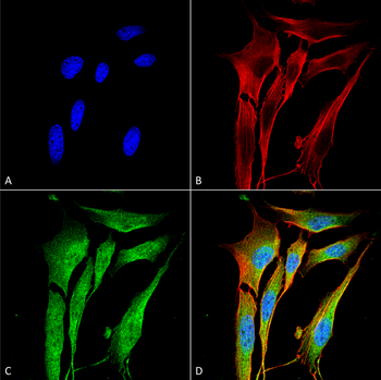
Immunocytochemistry/Immunofluorescence analysis using Mouse Anti-VAChT Monoclonal Antibody, Clone N6/38. Tissue: Neuroblastoma cell line (SK-N-BE). Species: Human. Fixation: 4% Formaldehyde for 15 min at RT. Primary Antibody: Mouse Anti-VAChT Monoclonal Antibody at 1:100 for 60 min at RT. Secondary Antibody: Goat Anti-Mouse ATTO 488 at 1:200 for 60 min at RT. Counterstain: Phalloidin Texas Red F-Actin stain; DAPI (blue) nuclear stain at 1:1000, 1:5000 for 60 min at RT, 5 min at RT. Localization: Membrane. Magnification: 60X. (A) DAPI (blue) nuclear stain. (B) Phalloidin Texas Red F-Actin stain. (C) VAChT Antibody. (D) Composite.
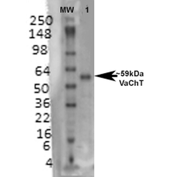
Western Blot analysis of Rat brain membrane lysate showing detection of VAChT protein using Mouse Anti-VAChT Monoclonal Antibody, Clone N6/38. Primary Antibody: Mouse Anti-VAChT Monoclonal Antibody at 1:1000.

Vesicular Acetylcholine Transporter Rabbit Polyclonal Antibody (Biotin) [orb457889]
WB
Rabbit
Human, Mouse, Rat
Rabbit
Polyclonal
Biotin
100 μl
