You have no items in your shopping cart.
Cart summary
Item 1 of 6
Item 1 of 6
UCHL1 Antibody (C-term)
Catalog Number: orb1931713

| Catalog Number | orb1931713 |
|---|---|
| Category | Antibodies |
| Description | Purified Rabbit Polyclonal Antibody (Pab) |
| Species/Host | Rabbit |
| Clonality | Polyclonal |
| Clone Number | RB7379 |
| Tested applications | FC, IF, IHC-P, WB |
| Predicted Reactivity | Equine, Porcine |
| Reactivity | Human, Mouse, Rat |
| Isotype | Rabbit IgG |
| Dilution range | IF: 1:10~50, WB: 1:1000, IHC-P: 1:50~100, IHC-P: 1:25, IHC-P: 1:25, FC: 1:10~50 |
| Form/Appearance | Purified polyclonal antibody supplied in PBS with 0.09% (W/V) sodium azide. This antibody is purified through a protein A column, followed by peptide affinity purification. |
| Conjugation | Unconjugated |
| MW | 24824 Da |
| Target | This UCHL1 antibody is generated from rabbits immunized with a KLH conjugated synthetic peptide between 187-216 amino acids from the C-terminal region of human UCHL1. |
| UniProt ID | P09936 |
| NCBI | NP_004172.2 |
| Storage | Maintain refrigerated at 2-8°C for up to 2 weeks. For long term storage store at -20°C in small aliquots to prevent freeze-thaw cycles |
| Alternative names | Ubiquitin carboxyl-terminal hydrolase isozyme L1, Read more... |
| Note | For research use only |
| Expiration Date | 12 months from date of receipt. |

UCHL1 Antibody (C-term) flow cytometric analysis of NCI-H460 cells (right histogram) compared to a negative control cell (left histogram). FITC-conjugated goat-anti-rabbit secondary antibodies were used for the analysis.

Confocal immunofluorescent analysis of UCHL1 Antibody (C-term) with NCI-H460 cell followed by Alexa Fluor 488-conjugated goat anti-rabbit lgG (green). DAPI was used to stain the cell nuclear (blue).
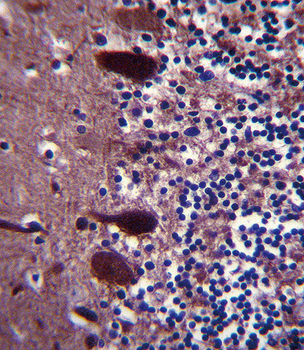
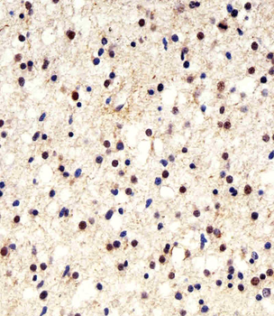
Staining UCHL1 in human brain tissue sections by Immunohistochemistry (IHC-P - paraformaldehyde-fixed, paraffin-embedded sections). Tissue was fixed with formaldehyde and blocked with 3% BSA for 0.5 hour at room temperature; antigen retrieval was by heat mediation with a citrate buffer (pH6). Samples were incubated with primary antibody (1/25) for 1 hours at 37°C. A undiluted biotinylated goat polyvalent antibody was used as the secondary antibody.
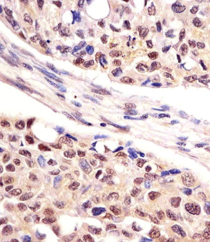
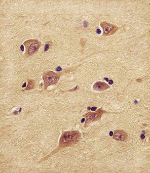
Staining UCHL1 in human lung adenocarcinoma tissue sections by Immunohistochemistry (IHC-P - paraformaldehyde-fixed, paraffin-embedded sections). Tissue was fixed with formaldehyde and blocked with 3% BSA for 0.5 hour at room temperature; antigen retrieval was by heat mediation with a citrate buffer (pH6). Samples were incubated with primary antibody (1/25) for 1 hours at 37°C. A undiluted biotinylated goat polyvalent antibody was used as the secondary antibody.
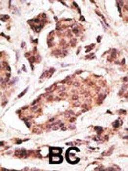
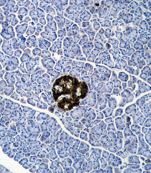
Formalin-fixed and paraffin-embedded human cancer tissue reacted with the primary antibody, which was peroxidase-conjugated to the secondary antibody, followed by DAB staining. This data demonstrates the use of this antibody for immunohistochemistry; clinical relevance has not been evaluated. BC = breast carcinoma; HC = hepatocarcinoma.
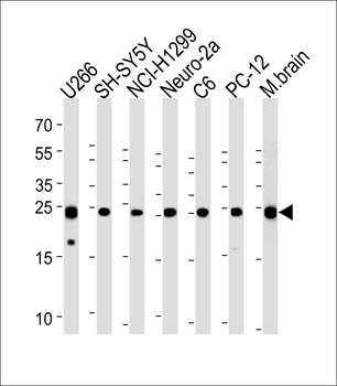
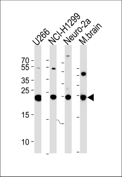
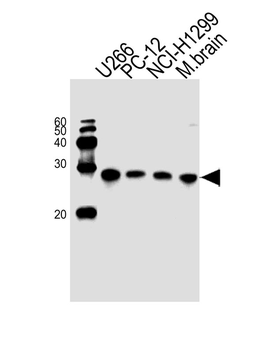
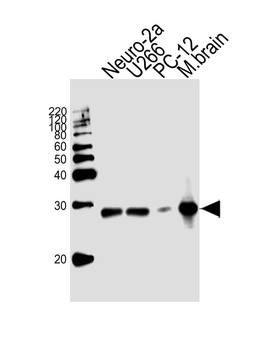
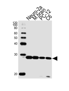
UCHL1 Antibody (C-term) western blot analysis in U266, SH-SY5Y, NCI-H1299, mouse Neuro-2a, rat C6, PC-12 cell line and mouse brain tissue lysates (35 ug/lane). This demonstrates the UCHL1 antibody detected the UCHL1 protein (arrow).
- Item 1 of 4
UCHL1 Antibody (C-term) [orb1931712]
IF, IHC-P, WB
Equine, Mouse, Porcine
Human, Rat
Rabbit
Polyclonal
Unconjugated
50 μl, 100 μl - Item 1 of 3
- Item 1 of 3
UCHL1 Antibody (C-term) [orb1927319]
IHC-P, WB
Equine, Mouse, Porcine
Human, Rat
Mouse
Monoclonal
Unconjugated
50 μl, 100 μl - Item 1 of 2
UCHL1 Antibody (C-term) [orb1788247]
WB
Equine, Porcine
Human, Mouse, Rat
Rabbit
Polyclonal
Unconjugated
100 μl - Item 1 of 1