You have no items in your shopping cart.
Cart summary
Item 1 of 4
Item 1 of 4
TrpV3 Antibody: FITC
Catalog Number: orb148706

| Catalog Number | orb148706 |
|---|---|
| Category | Antibodies |
| Description | Mouse monoclonal to TrpV3 (FITC). The TRPV3 protein belongs to a family of non-selective cation channels that function in a variety of processes, including temperature sensation and vasoregulation. The thermo sensitive members of this family are expressed in subsets of sensory neurons that terminate in the skin, and are activated at distinct physiological temperatures. This channel is activated at temperatures between 22 and 40 degrees C. The gene lies in close proximity to another family member (TRPV1) gene on chromosome 17, and the two encoded proteins are thought to associate with each other to form heteromeric channels. |
| Species/Host | Mouse |
| Clonality | Monoclonal |
| Clone Number | N15/39 (Formerly sold as S15-39) |
| Tested applications | ICC, IF, IHC |
| Reactivity | Human, Mouse, Rat |
| Isotype | IgG2a |
| Immunogen | Synthetic peptide amino acids 458-474 (cytoplasmic C-terminus) of rat TrpV3 |
| Concentration | 1 mg/ml |
| Dilution range | WB (1:1000), IHC (1:1000), ICC/IF (1:100) |
| Conjugation | FITC |
| MW | 70kDa |
| Target | TRPV3 |
| Entrez | 497948 |
| UniProt ID | Q4QYD9 |
| NCBI | NP_001020928 |
| Storage | Conjugated antibodies should be stored according to the product label |
| Buffer/Preservatives | 640.91mM DMSO, 136.36mM Ethanolamine, 9.09mM Sodium Bicarbonate in 90.9% PBS |
| Alternative names | VRL3 antibody, vanilloid receptor 2 antibody, tran Read more... |
| Note | For research use only |
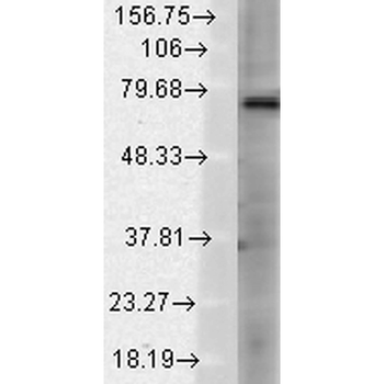
| Application notes | 1 µg/ml of SMC-334 was sufficient for detection of TrpV3 in 10 µg of COS-1 cell lysate transiently transfected with TrpV3 by colorimetric immunoblot analysis using Goat anti-mouse IgG:HRP as the secondary antibody |
| Expiration Date | 12 months from date of receipt. |

Immunocytochemistry/Immunofluorescence analysis using Mouse Anti-TrpV3 Monoclonal Antibody, Clone S15-39. Tissue: Neuroblastoma cells (SH-SY5Y). Species: Human. Fixation: 4% PFA for 15 min. Primary Antibody: Mouse Anti-TrpV3 Monoclonal Antibody at 1:50 for overnight at 4°C with slow rocking. Secondary Antibody: AlexaFluor 488 at 1:1000 for 1 hour at RT. Counterstain: Phalloidin-iFluor 647 (red) F-Actin stain; Hoechst (blue) nuclear stain at 1:800, 1.6mM for 20 min at RT. (A) Hoechst (blue) nuclear stain. (B) Phalloidin-iFluor 647 (red) F-Actin stain. (C) TrpV3 Antibody (D) Composite.

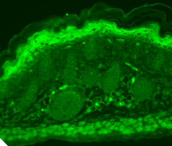
Immunohistochemistry analysis using Mouse Anti-TrpV3 Monoclonal Antibody, Clone S15-39. Tissue: backskin. Species: Mouse. Fixation: Bouin's Fixative and paraffin-embedded. Primary Antibody: Mouse Anti-TrpV3 Monoclonal Antibody at 1:100 for 1 hour at RT. Secondary Antibody: FITC Goat Anti-Mouse (green) at 1:50 for 1 hour at RT. Localization: Filaggrin-like staining in upper layers. Dull lower layer cell staining. Some stain seen in hypodermis.
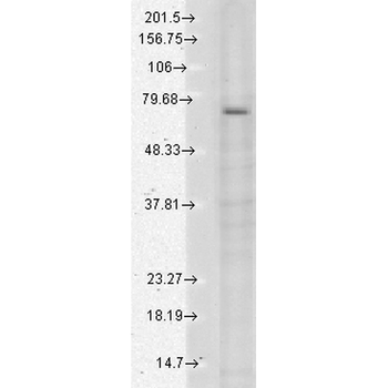
Western Blot analysis of Rat brain membrane lysate showing detection of TrpV3 protein using Mouse Anti-TrpV3 Monoclonal Antibody, Clone S15-39. Load: 15 μg. Block: 1.5% BSA for 30 minutes at RT. Primary Antibody: Mouse Anti-TrpV3 Monoclonal Antibody at 1:1000 for 2 hours at RT. Secondary Antibody: Sheep Anti-Mouse IgG: HRP for 1 hour at RT.
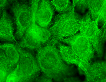
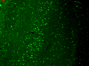
Immunocytochemistry/Immunofluorescence analysis using Mouse Anti-TrpV3 Monoclonal Antibody, Clone S15-39. Tissue: HaCaT cells. Species: Human. Fixation: Cold 100% methanol for 10 minutes at -20°C. Primary Antibody: Mouse Anti-TrpV3 Monoclonal Antibody at 1:100 for 1 hour at RT. Secondary Antibody: FITC Goat Anti-Mouse (green) at 1:50 for 1 hour at RT. Localization: Dotty staining in all cells. Some intermediate filament-like staining in some cells.
- Item 1 of 4