You have no items in your shopping cart.
Cart summary
Item 1 of 4
Item 1 of 4
TrpV3 Antibody: Biotin
Catalog Number: orb148484

| Catalog Number | orb148484 |
|---|---|
| Category | Antibodies |
| Description | Mouse monoclonal to TrpV3 (Biotin). Ion channels are integral membrane proteins that help establish and control the small voltage gradient across the plasma membrane of living cells by allowing the flow of ions down their electrochemical gradient. They are present in the membranes that surround all biological cells because their main function is to regulate the flow of ions across this membrane. Whereas some ion channels permit the passage of ions based on charge, others conduct based on a ionic species, such as sodium or potassium. Furthermore, in some ion channels, the passage is governed by a gate which is controlled by chemical or electrical signals, temperature, or mechanical forces. There are a few main classifications of gated ion channels. There are voltage- gated ion channels, ligand-gated, other gating systems and finally those that are classified differently, having more exotic characteristics. The first are voltage- gated ion channels which open and close in response to membrane potential. These are then separated into sodium, calcium, potassium, proton, transient receptor, and cyclic nucleotide-gated channels; each of which is responsible for a unique role. Ligand-gated ion channels are also known as ionotropic receptors, and they open in response to specific ligand molecules binding to the extracellular domain of the receptor protein. The other gated classifications include activation and inactivation by second messengers, inward-rectifier potassium channels, calcium-activated potassium channels, two-pore-domain potassium channels, light-gated channels, mechano-sensitive ion channels and cyclic nucleotide-gated channels. Finally, the other classifications are based on less normal characteristics such as two-pore channels, and transient receptor potential channels. The TRPV3 protein belongs to a family of nonselective cation channels that function in a variety of processes, including temperature sensation and vasoregulation. The thermosensitive members of this family are expressed in subsets of sensory neurons that terminate in the skin, and are activated at distinct physiological temperatures. This channel is activated at temperatures between 22 and 40 degrees C. The gene lies in close proximity to another family member (TRPV1) gene on chromosome 17, and the two encoded proteins are thought to associate with each other to form heteromeric channels... |
| Species/Host | Mouse |
| Clonality | Monoclonal |
| Clone Number | N15/4 (Formerly sold as S15-4) |
| Tested applications | ELISA, ICC, IF, IHC, WB |
| Reactivity | Human, Mouse, Rat |
| Isotype | IgG2a |
| Immunogen | Synthetic peptide amino acids 774-791 (C-terminus) of rat TrpV3 |
| Concentration | 1 mg/ml |
| Dilution range | WB (1:1000), IHC (1:1000), ICC/IF (1:100) |
| Conjugation | Biotin |
| MW | 70kDa |
| Target | TRPV3 |
| Entrez | 497948 |
| UniProt ID | Q4QYD9 |
| NCBI | NP_001020928 |
| Storage | Conjugated antibodies should be stored according to the product label |
| Buffer/Preservatives | 136.36mM Ethanolamine, and 9.55mM Sodium Bicarbonate in 95.45% PBS |
| Alternative names | 1110036I10Rik antibody, MGC124324 antibody, MGC124 Read more... |
| Note | For research use only |
| Application notes | 1 µg/ml of SMC-319 was sufficient for detection of TrpV3 in 10 µg of COS-1 cell lysate transiently transfected with TrpV3 by colorimetric immunoblot analysis using Goat anti-mouse IgG:HRP as the secondary antibody. |
| Expiration Date | 12 months from date of receipt. |

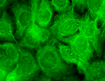
Immunocytochemistry/Immunofluorescence analysis using Mouse Anti-TrpV3 Monoclonal Antibody, Clone N15/4. Tissue: Neuroblastoma cells (SH-SY5Y). Species: Human. Fixation: 4% PFA for 15 min. Primary Antibody: Mouse Anti-TrpV3 Monoclonal Antibody at 1:100 for overnight at 4°C with slow rocking. Secondary Antibody: AlexaFluor 488 at 1:1000 for 1 hour at RT. Counterstain: Phalloidin-iFluor 647 (red) F-Actin stain; Hoechst (blue) nuclear stain at 1:800, 1.6mM for 20 min at RT. (A) Hoechst (blue) nuclear stain. (B) Phalloidin-iFluor 647 (red) F-Actin stain. (C) TrpV3 Antibody (D) Composite.
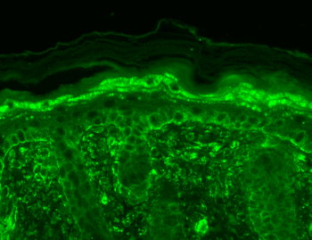
Immunohistochemistry analysis using Mouse Anti-TrpV3 Monoclonal Antibody, Clone N15/4. Tissue: backskin. Species: Mouse. Fixation: Bouin's Fixative and paraffin-embedded. Primary Antibody: Mouse Anti-TrpV3 Monoclonal Antibody at 1:100 for 1 hour at RT. Secondary Antibody: FITC Goat Anti-Mouse (green) at 1:50 for 1 hour at RT.
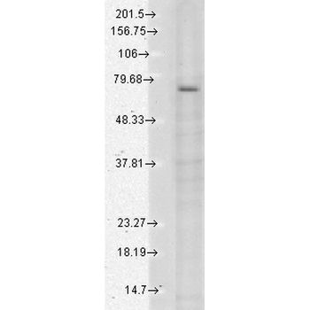
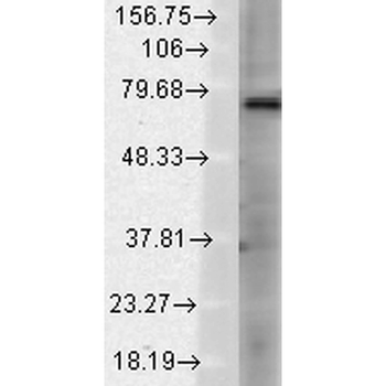
Western Blot analysis of Human Cell lysates showing detection of TrpV3 protein using Mouse Anti-TrpV3 Monoclonal Antibody, Clone N15/4. Load: 15 μg. Block: 1.5% BSA for 30 minutes at RT. Primary Antibody: Mouse Anti-TrpV3 Monoclonal Antibody at 1:1000 for 2 hours at RT. Secondary Antibody: Sheep Anti-Mouse IgG: HRP for 1 hour at RT.
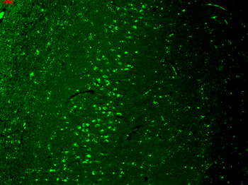
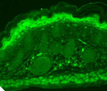
Immunohistochemistry analysis using Mouse Anti-TrpV3 Monoclonal Antibody, Clone N15/4. Tissue: hippocampus. Species: Human. Fixation: Bouin's Fixative and paraffin-embedded. Primary Antibody: Mouse Anti-TrpV3 Monoclonal Antibody at 1:1000 for 1 hour at RT. Secondary Antibody: FITC Goat Anti-Mouse (green) at 1:50 for 1 hour at RT.
- Item 1 of 4
TrpV3 Antibody: Biotin [orb148705]
ELISA, ICC, IF, IHC, WB
Human, Mouse, Rat
Mouse
Monoclonal
Biotin
100 μg