You have no items in your shopping cart.
Cart summary
Item 1 of 4
Item 1 of 4
TRPC6 antibody
Catalog Number: orb344463

| Catalog Number | orb344463 |
|---|---|
| Category | Antibodies |
| Description | TRPC6 antibody |
| Species/Host | Mouse |
| Clonality | Monoclonal |
| Clone Number | 3F2.H10.F2 |
| Tested applications | ELISA, IHC, WB |
| Reactivity | Human, Mouse |
| Isotype | IgG1 |
| Immunogen | This monoclonal antibody was produced by repeated immunizations with a synthetic peptide corresponding to a region near the carboxy terminus of human TRPC6 protein. |
| Concentration | 1.0 mg/mL |
| Dilution range | ELISA: 1:10,000 - 1:50,000, IHC: 2.5 µg/mL, WB: 1:500- 1:2,000 |
| Form/Appearance | Liquid (sterile filtered) |
| Purity | This product was purified from concentrated tissue culture supernate by Protein A chromatography. This antibody is specific for human TRPC6 protein. A BLAST analysis was used to suggest cross-reactivity with TRPC6 from chimpanzee based on 100% homology with the immunizing sequence. Cross-reactivity with TRPC6 from other sources has not been determined. |
| Conjugation | Unconjugated |
| UniProt ID | Q9Y210 |
| NCBI | 5730102 |
| Storage | Store vial at -20° C or below prior to opening. This vial contains a relatively low volume of reagent (25 µL). To minimize loss of volume dilute 1:10 by adding 225 µL of the buffer stated above directly to the vial. Recap, mix thoroughly and briefly centrifuge to collect the volume at the bottom of the vial. Use this intermediate dilution when calculating final dilutions as recommended below. Store the vial at -20°C or below after dilution. Avoid cycles of freezing and thawing. |
| Buffer/Preservatives | 0.01% (w/v) Sodium Azide |
| Alternative names | mouse anti-TRPC6 Antibody, TRPC 6, TRP6, short tra Read more... |
| Note | For research use only |
| Application notes | Anti-TRPC6 monoclonal antibody has been tested by ELISA, immunohistochemistry and western blotting. Expect a band approximately 30 kDa in size corresponding to the cytoplasmic domain of TRPC6 protein by western blotting in the appropriate cell lysate or extract. Specific conditions for reactivity should be optimized by the end user. Use formalin-fixed paraffin-embedded sections for immunohistochemistry. No pre-treatment of sample is required. Strong staining was observed in adrenal, Purkinje neurons, cortical neurons, heart, ganglion cells, renal tubules, Sertoli cells, hepatocytes, skeletal muscle, exocrine pancreas, and germinal centers of lymphoid follicles. Moderate staining was observed in colon epithelium and plasma cells, B-lymphocytes, and parafollicular cells of the thyroid. Faint staining was seen in respiratory epithelium. Prostate and placenta were negative for staining. The antibody produced minimal to no background staining and appeared very specific at 2.5 µg/mL. |
| Expiration Date | 12 months from date of receipt. |

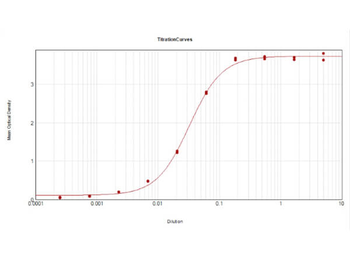
ELISA Results of Mouse Anti-TRPC6 Antibody. Each well was coated in duplicate with 0.1 µg of conjugate. The working dilution is 1:31000. The starting dilution of antibody was 5 µg/ml and the X-axis represents the Log10 of a 3-fold dilution. This titration is a 4-parameter curve fit where the IC50 is defined as the titer of the antibody. Assay performed using HRP conjugation Stabilizer, Rabbit Anti-Mouse IgG HRP conjugated (p/n orb347506) and TMB substrate (p/n orb348651).

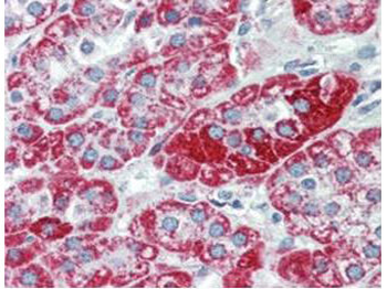
Immunohistochemistry using Biorbyt's anti-TRPC6 monoclonal antibody shows detection of TRPC6 in human adrenal (cortex) tissue (40X). The antibody was used a dilution to 2.5 µg/mL. The image shows strong staining with minimal background staining. Tissue was formalin fixed and paraffin embedded. No pre-treatment of sample was required. The image shows the localization of antibody as the precipitated red signal, with a hematoxylin purple nuclear counterstain.

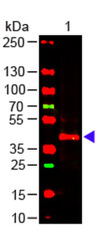
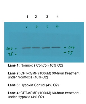
Western Blot of Mouse anti-TRPC6 Antibody. Lane 1: Mouse Kidney WCL (p/n orb348717). Load: 10 µg per lane. Primary antibody: TRPC6 Antibody at 1:1000 for overnight at 4°C. Secondary antibody: donkey anti-mouse DyLight™ 649 at 1:20000 for 30 min at RT. Block: orb348637 for 30 min at RT.
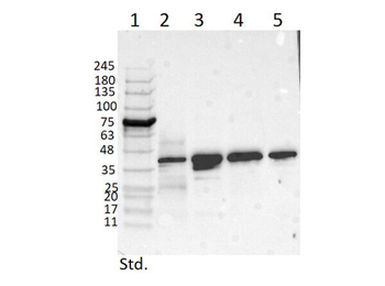
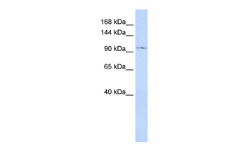
Western Blot of Mouse Anti-TRPC6 Antibody. Lane 1: Opal Prestained Molecular Weight Marker. Lane 2: Mouse Pancreas Tissue Lysate (p/n orb348719) [10 µg]. Lane 3: MCF-7 Whole Cell Lysate (p/n orb348664) [10 µg]. Lane 4: A431 Whole Cell Lysate (p/n orb348665) [10 µg]. Lane 5: Jurkat Whole Cell Lysate (p/n orb348674) [10 µg]. Primary Antibody: Anti-TRPC6 at 1 µg/mL overnight at 2-8°C. Secondary Antibody: Rabbit Anti-Mouse IgG Peroxidase (p/n orb347506) 1:40000 for 30 mins at RT. Blocking Buffer: BlockOut Buffer (p/n orb348644) for 30 mins at RT. Predicted MW: ~30 kDa. Observed MW: ~40 kDa. Exposure: 5sec.
- Item 1 of 4
TRPC6 Antibody [orb1238603]
ELISA, IF, IHC-P, WB
Human, Mouse
Rabbit
Polyclonal
Unconjugated
0.1 mg, 0.02 mg - Item 1 of 4
TRPC6 Antibody [orb1238605]
ELISA, IF, IHC-P, WB
Human, Mouse, Rat
Rabbit
Polyclonal
Unconjugated
0.1 mg, 0.02 mg - Item 1 of 4
- Item 1 of 4
TRPC6 antibody [orb329813]
WB
Animal, Bovine, Canine, Guinea pig, Human, Mouse, Porcine, Rat, Yeast
Canine, Equine, Guinea pig, Human, Mouse, Porcine, Rat, Yeast
Rabbit
Polyclonal
Unconjugated
100 μl - Item 1 of 2


Submit a review
Filter by Rating

- 5 stars

- 4 stars
- 3 stars
- 2 stars
- 1 stars
