You have no items in your shopping cart.
Cart summary
Item 1 of 4
Item 1 of 4
TNFRSF6B Antibody (N-term)
Catalog Number: orb1939148

| Catalog Number | orb1939148 |
|---|---|
| Category | Antibodies |
| Description | Affinity Purified Rabbit Polyclonal Antibody (Pab) |
| Species/Host | Rabbit |
| Clonality | Polyclonal |
| Clone Number | RB21898 |
| Tested applications | FC, IHC-P, WB |
| Reactivity | Human |
| Isotype | Rabbit IgG |
| Dilution range | WB: 1:2000, WB: 1:1000, IHC-P: 1:50~100, FC: 1:10~50 |
| Form/Appearance | Purified polyclonal antibody supplied in PBS with 0.09% (W/V) sodium azide. This antibody is purified through a protein A column, followed by peptide affinity purification. |
| Conjugation | Unconjugated |
| MW | 32680 Da |
| Target | This TNFRSF6B antibody is generated from rabbits immunized with a KLH conjugated synthetic peptide between 22-48 amino acids from the N-terminal region of human TNFRSF6B. |
| UniProt ID | O95407 |
| NCBI | NP_003814.1 |
| Storage | Maintain refrigerated at 2-8°C for up to 2 weeks. For long term storage store at -20°C in small aliquots to prevent freeze-thaw cycles |
| Alternative names | Tumor necrosis factor receptor superfamily member Read more... |
| Note | For research use only |
| Expiration Date | 12 months from date of receipt. |

All lanes: Anti-TNFRSF6B Antibody (N-term) at 1:2000 dilution. Lane 1: Hela whole cell lysate. Lane 2: HCT116 whole cell lysate. Lane 3: PC-3 whole cell lysate. Lane 4: SW480 whole cell lysate. Lane 5: HUVEC whole cell lysate. Lysates/proteins at 20 µg per lane. Secondary Goat Anti-Rabbit IgG, (H+L), Peroxidase conjugated at 1/10000 dilution. Predicted band size: 33 kDa. Blocking/Dilution buffer: 5% NFDM/TBST.
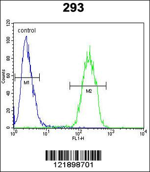
TNFRSF6B Antibody (N-term) flow cytometric analysis of 293 cells (right histogram) compared to a negative control (rabbit IgG alone) (left histogram). FITC-conjugated goat-anti-rabbit secondary antibodies were used for the analysis.

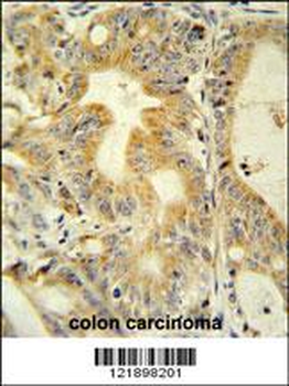
TNFRSF6B antibody (N-term) immunohistochemistry analysis in formalin fixed and paraffin embedded human colon carcinoma followed by peroxidase conjugation of the secondary antibody and DAB staining. This data demonstrates the use of the TNFRSF6B antibody (N-term) for immunohistochemistry. Clinical relevance has not been evaluated.

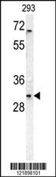
TNFRSF6B Antibody (N-term) western blot analysis in 293 cell line lysates (35 ug/lane). This demonstrates the TNFRSF6B antibody detected the TNFRSF6B protein (arrow).
- Item 1 of 4