You have no items in your shopping cart.
Cart summary
Item 1 of 6
Item 1 of 6
TNF-R1 Antibody: Biotin
Catalog Number: orb151862

| Catalog Number | orb151862 |
|---|---|
| Category | Antibodies |
| Description | Rabbit polyclonal antibody against TNFR1 conjugated to Biotin |
| Species/Host | Rabbit |
| Clonality | Polyclonal |
| Tested applications | ELISA, ICC, IF, IHC, WB |
| Reactivity | Bovine, Canine, Human, Monkey, Mouse, Rabbit, Rat |
| Immunogen | Peptide corresponding to AA 20-43 of the mouse TNF-R1 sequence, identical to rat and human over those residues |
| Concentration | 1 mg/ml |
| Dilution range | WB (1:1000), IHC (1:100), ICC/IF (1:100) |
| Conjugation | Biotin |
| MW | 55kDa |
| Target | TNFR1 |
| Entrez | 8666 |
| UniProt ID | P19438 |
| NCBI | P19438 |
| Storage | Conjugated antibodies should be stored according to the product label |
| Buffer/Preservatives | 136.36mM Ethanolamine, and 9.55mM Sodium Bicarbonate in 95.45% PBS |
| Alternative names | Tumor necrosis factor receptor 1 antibody, TNFR-1 Read more... |
| Note | For research use only |
| Application notes | 1 µg/ml of SPC-170 was sufficient for detection of TNFR1 in 20 µg of Hela lysate by colorimetric immunoblot analysis using Goat anti-rabbit IgG:HRP as the secondary antibody. |
| Expiration Date | 12 months from date of receipt. |

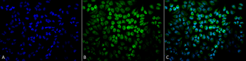
Immunocytochemistry/Immunofluorescence analysis using Rabbit Anti-TNF-R1 Polyclonal Antibody. Tissue: Cervical cancer cell line (HeLa). Species: Human. Fixation: 2% Formaldehyde for 20 min at RT. Primary Antibody: Rabbit Anti-TNF-R1 Polyclonal Antibody at 1:100 for 12 hours at 4°C. Secondary Antibody: APC Goat Anti-Rabbit (red) at 1:200 for 2 hours at RT. Counterstain: DAPI (blue) nuclear stain at 1:40000 for 2 hours at RT. Localization: Golgi apparatus membrane. Magnification: 100x. (A) DAPI (blue) nuclear stain. (B) Anti-TNF-R1 Antibody. (C) Composite.
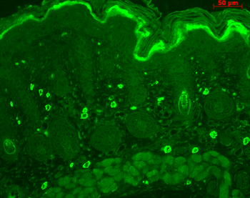
Immunohistochemistry analysis using Rabbit Anti-TNF-R1 Polyclonal Antibody. Tissue: backskin. Species: Mouse. Fixation: Bouin's Fixative Solution. Primary Antibody: Rabbit Anti-TNF-R1 Polyclonal Antibody at 1:100 for 1 hour at RT. Secondary Antibody: FITC Goat Anti-Rabbit (green) at 1:50 for 1 hour at RT. Localization: dermis.
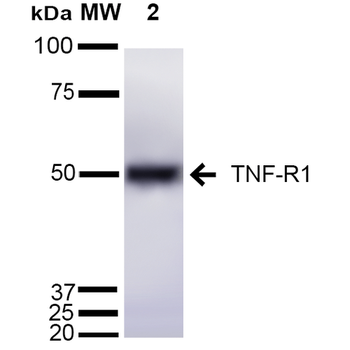
Western blot analysis of Mouse Liver cell lysates showing detection of ~55 kDa TNF-R1 protein using Rabbit Anti-TNF-R1 Polyclonal Antibody. Lane 1: Molecular Weight Ladder (MW). Lane 2: Mouse Liver cell lysates. Load: 15 μg. Block: 5% Skim Milk in 1X TBST. Primary Antibody: Rabbit Anti-TNF-R1 Polyclonal Antibody at 1:1000 for 2 hours at RT. Secondary Antibody: Goat Anti-Rabbit IgG: HRP at 1:2000 for 60 min at RT. Color Development: ECL solution for 5 min at RT. Predicted/Observed Size: ~55 kDa.
Immunocytochemistry/Immunofluorescence analysis using Rabbit Anti-TNF-R1 Polyclonal Antibody. Tissue: Cervical cancer cell line (HeLa). Species: Human. Fixation: 2% Formaldehyde for 20 min at RT. Primary Antibody: Rabbit Anti-TNF-R1 Polyclonal Antibody at 1:100 for 12 hours at 4°C. Secondary Antibody: FITC Goat Anti-Rabbit (green) at 1:200 for 2 hours at RT. Counterstain: DAPI (blue) nuclear stain at 1:40000 for 2 hours at RT. Localization: Golgi apparatus membrane. Magnification: 20x. (A) DAPI (blue) nuclear stain. (B) Anti-TNF-R1 Antibody. (C) Composite.
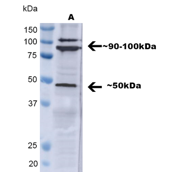
Western blot analysis of Human A549 showing detection of ~ 50 kDa TNF-R1 protein using Rabbit Anti-TNF-R1 Polyclonal Antibody. Lane 1: MW Ladder, Lane 2: A549. Load: 30 ug. Block: 5% BSA in TBST. Primary Antibody: Rabbit Anti-TNF-R1 Polyclonal Antibody at 1:1000 for 2 hours at RT with shaking. Secondary Antibody: Goat Anti-Rabbit IgG: HRP at 1:4000 for 1 hour at RT with shaking. Color Development: Chemiluminescent for HRP (Moss) for 5 min in RT. Predicted/Observed Size: ~ 50 kDa. Other Band (s): ~90-100kDa. Other bands can be explained by a few factors, such as oligomerization, self-aggregation, cleavage of the TNFR1 extracellular domain, etc.
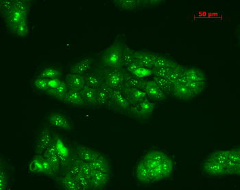
Immunocytochemistry/Immunofluorescence analysis using Rabbit Anti-TNF-R1 Polyclonal Antibody. Tissue: HaCaT cells. Species: Human. Fixation: Cold 100% methanol at -20°C for 10 minutes. Primary Antibody: Rabbit Anti-TNF-R1 Polyclonal Antibody at 1:100 for 12 hours at 4°C. Secondary Antibody: FITC Goat Anti-Rabbit at 1:50 for 1-2 hours at RT in dark. Localization: Punctate nuclear staining, dotty staining in cytoplasm.

- Item 1 of 1
TNFR1 Rabbit Polyclonal Antibody (Biotin) [orb1604201]
IF, IHC-Fr, IHC-P, WB
Human
Rabbit
Polyclonal
Biotin
100 μlTNFR1 Rabbit Polyclonal Antibody (Biotin) [orb458810]
FC, WB
Bovine, Canine, Equine, Porcine, Rabbit
Human, Mouse, Rat
Rabbit
Polyclonal
Biotin
100 μl

