You have no items in your shopping cart.
Cart summary
Item 1 of 4
Item 1 of 4
TNF Alpha antibody
Catalog Number: orb345126

| Catalog Number | orb345126 |
|---|---|
| Category | Antibodies |
| Description | TNF Alpha antibody |
| Species/Host | Rabbit |
| Clonality | Polyclonal |
| Tested applications | ELISA, IF, IHC, WB |
| Reactivity | Human |
| Isotype | IgG |
| Immunogen | The whole rabbit serum used to produce this IgG fraction antibody was prepared by repeated immunizations with recombinant human TNFα. |
| Concentration | 1.0 mg/mL |
| Dilution range | ELISA: 1:1,000 - 1:5,000, IHC: 1:100 - 1:500, IF: User Optimized, WB: 1:500 - 1:2,000 |
| Form/Appearance | Liquid (sterile filtered) |
| Purity | This antibody is primarily directed against mature 17,000 MW human TNFα and is useful in determining its presence in various assays. In general, this antibody also detects primate TNFα in the same formats using similar dilutions. The antibody does not recognize human TNFß (lymphotoxin). This IgG fraction antibody will recognize the cell-bound precursor of TNFα as a 26,000 protein in immunoblots, particularly in denatured samples. This antibody is also useful for neutralization of human and primate TNFα activity in bioassays. It does not neutralize the biological activity of lymphotoxin. For neutralization, it is recommended to incubate the sample with a 1:200 dilution of the antibody for at least 4 hours before being tested. A control of similarly diluted normal rabbit IgG is recommended. |
| Conjugation | Unconjugated |
| UniProt ID | P01375 |
| NCBI | P01375.1 |
| Storage | Store vial at -20° C or below prior to opening. This vial contains a relatively low volume of reagent (25 µL). To minimize loss of volume dilute 1:10 by adding 225 µL of the buffer stated above directly to the vial. Recap, mix thoroughly and briefly centrifuge to collect the volume at the bottom of the vial. Use this intermediate dilution when calculating final dilutions as recommended below. Store the vial at -20°C or below after dilution. Avoid cycles of freezing and thawing. |
| Buffer/Preservatives | None |
| Alternative names | APC1 antibody, Cachectin antibody, DIF antibody, D Read more... |
| Note | For research use only |
| Application notes | This IgG fraction antibody of anti-Human TNFα has been tested for use in neutralizations, ELISA, immunohistochemistry and immunoblotting. It recognizes the 17,000 MW TNFα. Reactivity in other immunoassays is unknown. |
| Expiration Date | 12 months from date of receipt. |
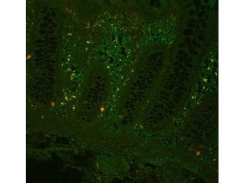
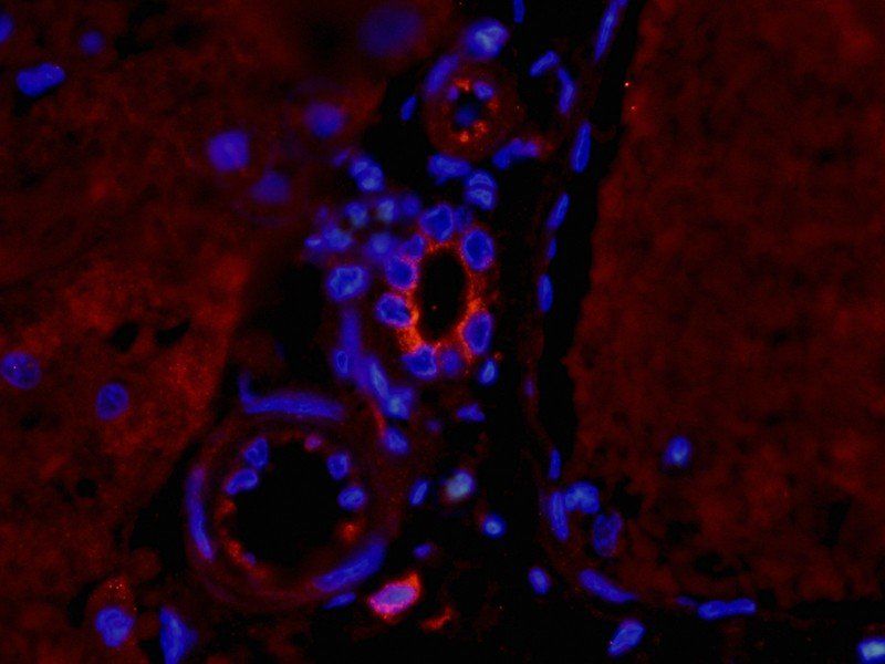
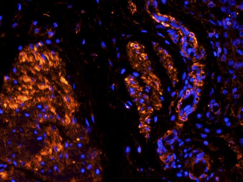
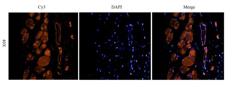
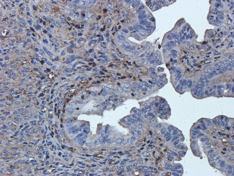
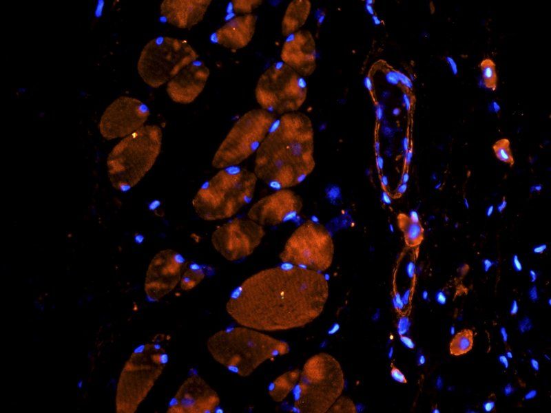
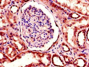
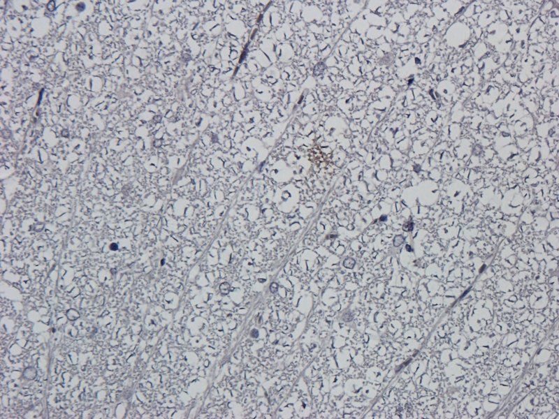
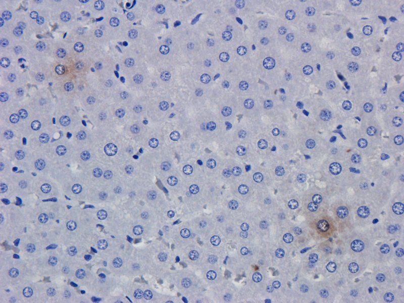
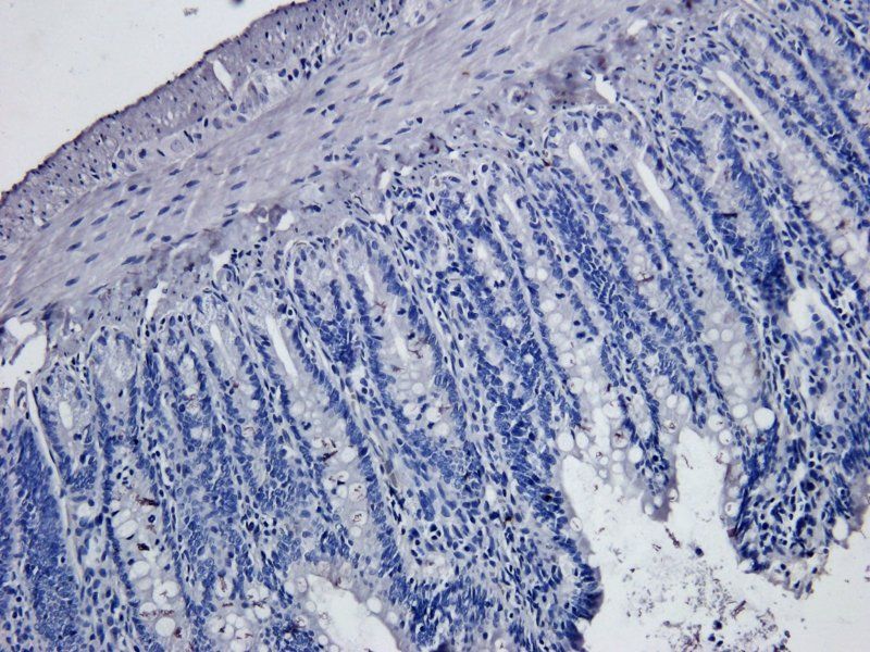
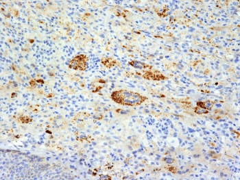
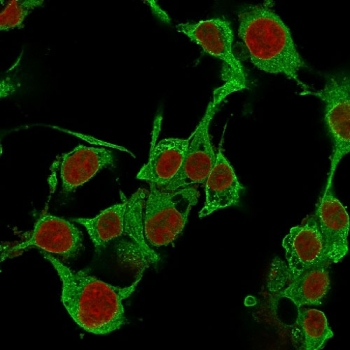
Fluorescent immunohistochemistry showing staining of human colon by Biorbyt's anti-TNF alpha (formalin/PFA-fixed paraffin-embedded sections). Samples were formaldehyde-fixed, then blocked in 10% serum for 2 hours at 20°C. The primary antibody was diluted 1:100 and incubated with the sample for 2 hours at 20°C. Alexa Fluor® 680 goat polyclonal secondary antibody was used diluted 1:5000.
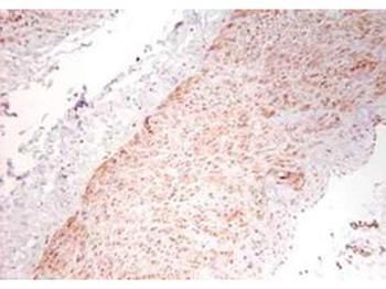
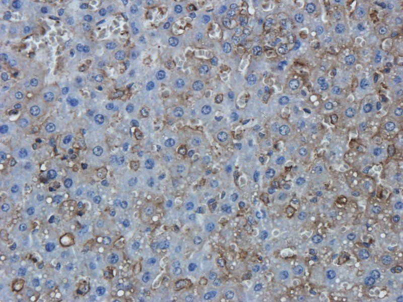
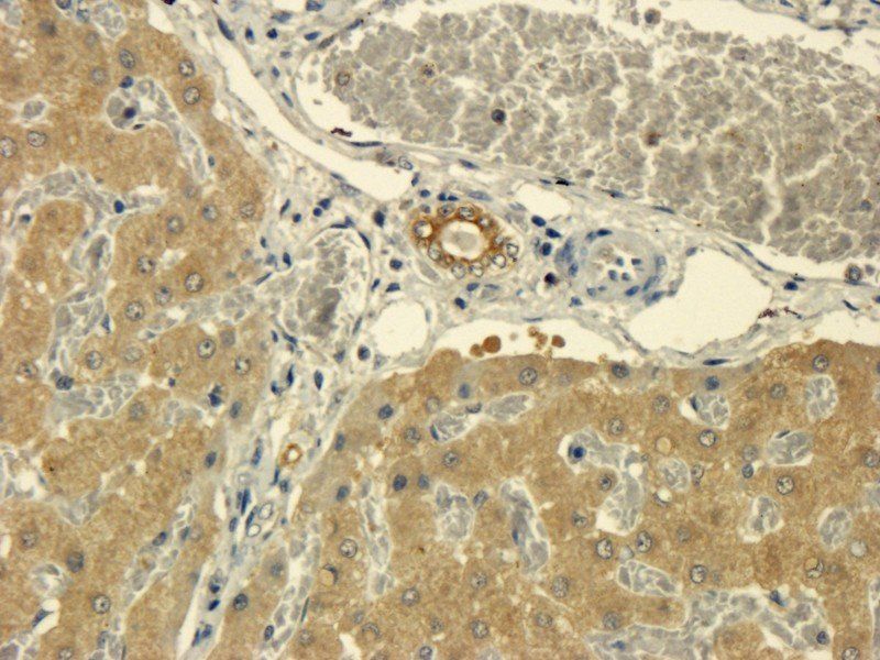
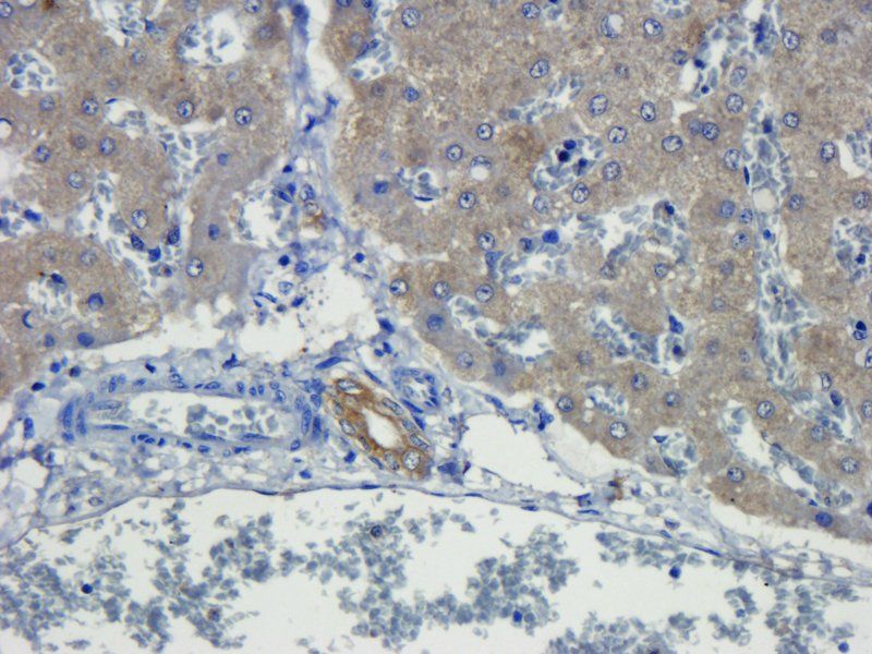
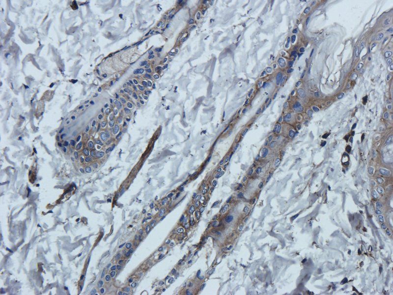
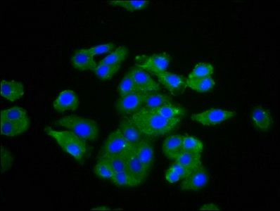
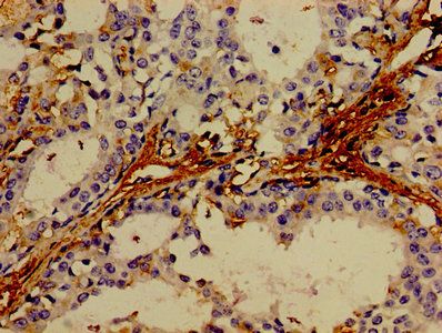
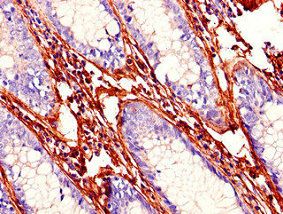
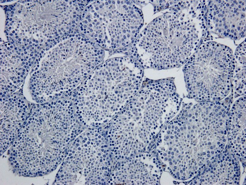
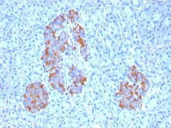
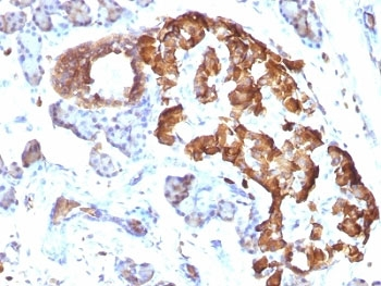
Immunohistochemistry using Biorbyt's polyclonal TNFa antibody showing staining of formalin/PFA-fixed paraffin-embedded sections of human artery tissue sections. Sections were fixed in formaldehyde and subjected to heat mediated antigen retrieval in citrate buffer (pH 6.0). Slides were blocked for ten minutes with 1.5% serum. Primary antibody was diluted 1:100 and incubated with samples for 24 hours at 4°C. HRP-conjugated goat anti-rabbit antibody was used as the secondary antibody.
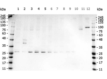
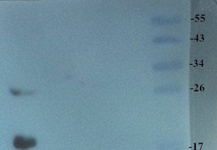
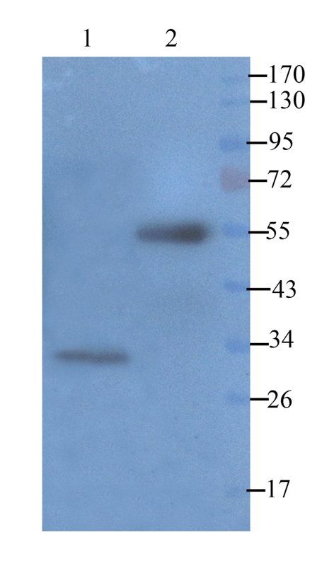
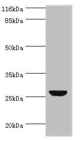
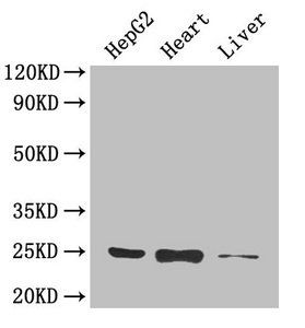
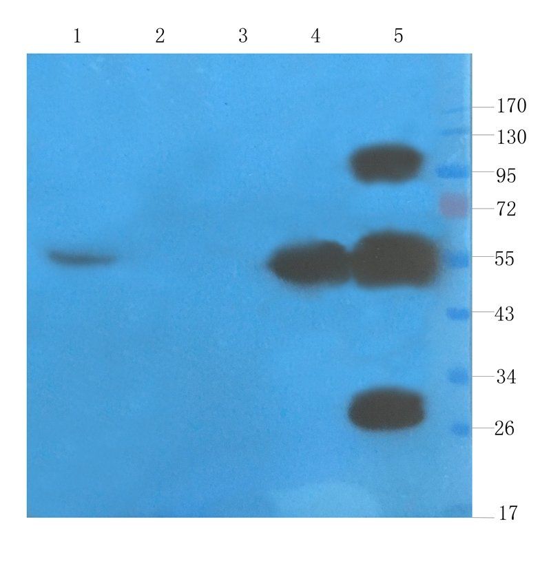
Western Blot of Rabbit anti-TNF Alpha antibody. Marker: Opal Pre-stained ladder. Lane 1: HEK293 lysate (p/n orb348669). Lane 2: HeLa Lysate (p/n orb348668). Lane 3: MCF-7 Lysate (p/n orb348664). Lane 4: Jurkat Lysate. Lane 5: A431 Lysate (p/n orb348665). Lane 6: A549 Lysate (p/n orb348675). Lane 7: LNCap Lysate (p/n orb348694). Lane 8: MOLT-4 Lysate (p/n orb348696). Lane 9: Ramos Lysate. Lane 10: Raji Lysate (p/n orb348672). Lane 11: A-172 Lysate (p/n orb348708). Lane 12: NIH/3T3 Lysate (p/n orb348714). Load: 35 µg per lane. Primary antibody: TNF Alpha antibody at 1 ug/ml overnight at 4C. Secondary antibody: Peroxidase rabbit secondary antibody (p/n orb347654) at 1:30000 for 60 min at RT. Blocking Buffer: 1% Casein-TTBS for 30 min at RT. Predicted/Observed size: 26 kDa for TNF Alpha.
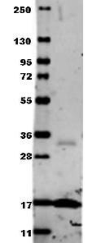
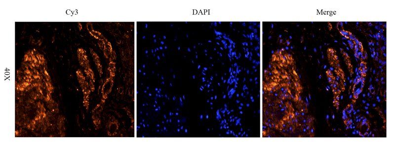
Western blot using Biorbyt's Anti-Human TNF-a (RABBIT) Antibody. Membrane blocked in 1% BSA-TBS-T for 30 min at RT, Rb-a-TNF alpha added at 1:1000 in 1% BSA-TBS-T o/n 4°C, DyLight 649 Gt-a-Rb added at 1:20000 in buffer (p/n orb348637) for 30 min at RT.
- Item 1 of 7
- Item 1 of 7
TNF alpha antibody [orb371962]
ICC, IF, IHC-P
Human, Mouse, Rat
Rabbit
Polyclonal
Unconjugated
200 μg, 100 μg - Item 1 of 6
Tumor necrosis factor antibody [orb239747]
ELISA, IF, IHC, WB
Human
Rabbit
Polyclonal
Unconjugated
50 μg, 100 μg - Item 1 of 6
- Item 1 of 6

Submit a review
Filter by Rating

- 5 stars

- 4 stars
- 3 stars
- 2 stars
- 1 stars
