You have no items in your shopping cart.
| Catalog Number | orb195994 |
|---|---|
| Category | Antibodies |
| Description | TIMP1 antibody |
| Species/Host | Rabbit |
| Clonality | Polyclonal |
| Tested applications | ELISA, ICC, IF, IHC-P, WB |
| Reactivity | Bovine, Canine, Guinea pig, Human, Mouse, Porcine, Rat, Sheep |
| Isotype | IgG |
| Immunogen | KLH conjugated synthetic peptide derived from human TIMP1. Please contact us for the exact immunogen sequence. The peptide is available as orb374835. |
| Concentration | - 100 μg (in 200 μl): 0.5 mg/ml- 200 μg (in 400 μl): 0.5 mg/ml |
| Dilution range | WB: 1:100-800, IHC-P: 1:400, IF/ICC: 1:400 |
| Form/Appearance | 10 mM PBS, 0.02% sodium azide |
| Purity | Polyclonal antibodies are purified by peptide affinity chromatography |
| Conjugation | Unconjugated |
| Target | TIMP1 |
| Entrez | 7076 |
| UniProt ID | P01033, P30120, P12032 |
| NCBI | 003254, 11, 003245, 21 |
| Storage | Maintain refrigerated at 2-8°C for up to 2 weeks. For long term storage store at -20°C in small aliquots to prevent freeze-thaw cycles. |
| Alternative names | anti Clgi antibody, anti Collagenase inhibitor ant Read more... |
| Note | For research use only |
| Expiration Date | 12 months from date of receipt. |
Filter by Reactivity
Carolina Brioschi Mathias a , Rebeca Ferreira Badaró b , Willian Grassi Bautz c , Leticia Nogueira da Gama-de-Souza HOW MALOCCLUSION INTERFERES WITH TISSUE INHIBITOR OF METALLOPROTEINASE-1 EXPRESSION AND MORPHOLOGY OF THE ARTICULAR CARTILAGE OF THE MANDIBLE IN FEMALE RATS Archives of Oral Biology, (2024)
Reactivity
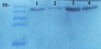
WB analysis of human breast cancer 4 (lane 1)
breast cancer 3 (lane 2), breast cancer 2 (lane 3), breast cancer 1 (lane 4) tissue using anti-TIMP1 (dilution of primary antibody - 1:100)
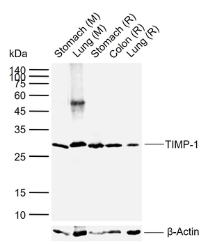
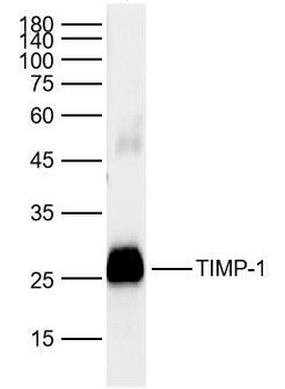
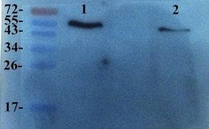
Western blot analysis of rat rat uterus(lane 1), rat kidney (lane 2) tissue using TIMP1 antibody (primary antibody at 1:300)
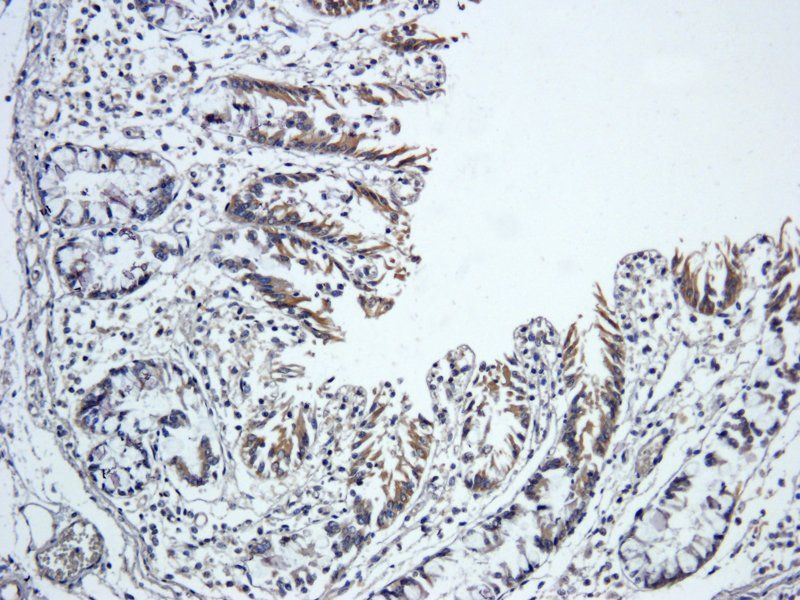
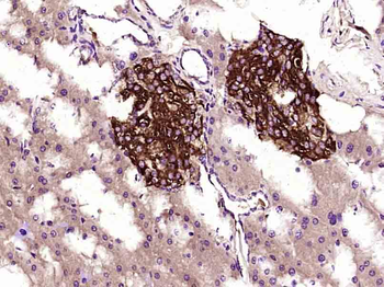
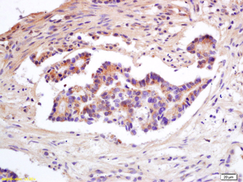
Immunohistochemical staining of paraffin embedded pig large intestine tissue using TIMP1 antibody (primary antibody at 1:400)
Immunohistochemical staining of paraffin embedded pig large intestine tissue using TIMP1 antibody (primary antibody at 1:400)
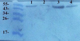
Western blot analysis of human breast cancer 4 (lane 1)
breast cancer 3 (lane 2), breast cancer 2 (lane 3), breast cancer 1 (lane 4) tissue using TIMP1 antibody (primary antibody at 1:200)
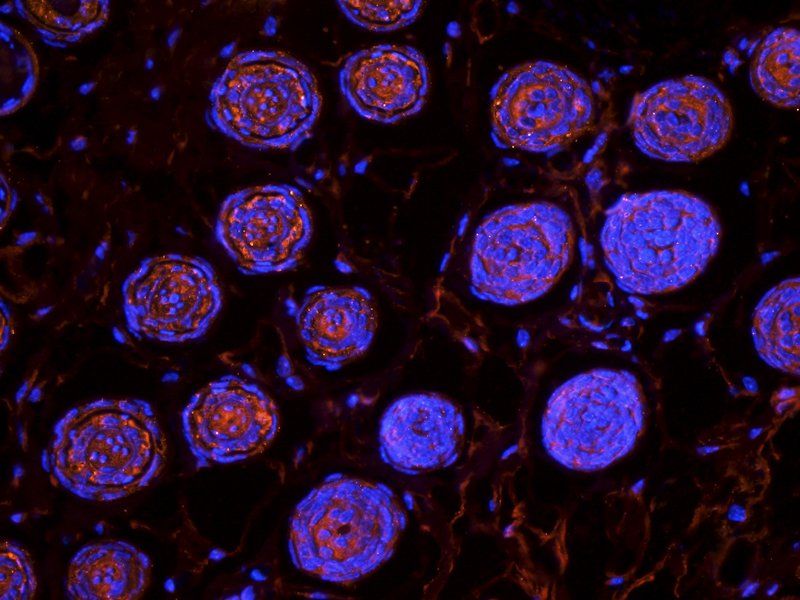
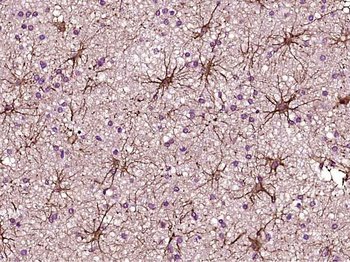
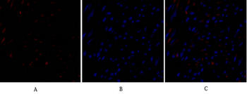
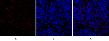
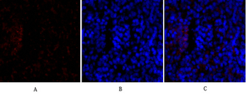
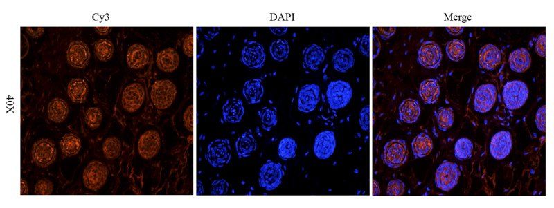
Immunofluorescence image of mouse skin tissue using TIMP1 antibody (dilution at 1:400)
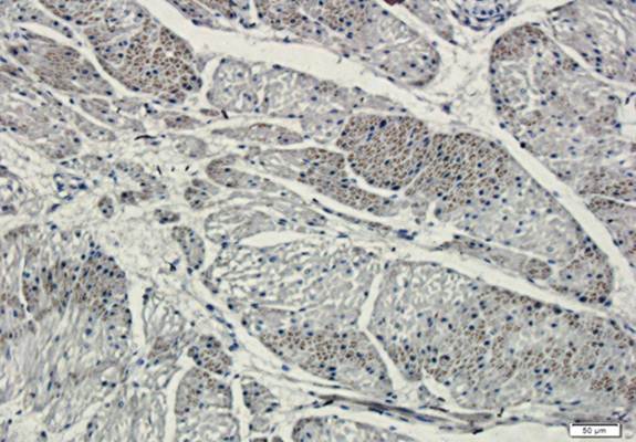
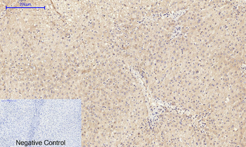
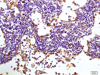
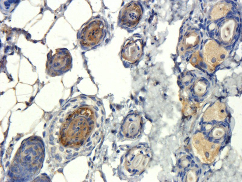
IHC-P image of mouse skin tissue using TIMP1 antibody (dilution of primary antibody at 1:400)
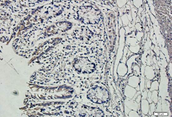
IF image of mouse skin tissue using anti-TIMP1 (primary antibody at 1:400)
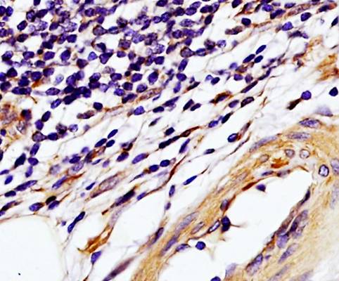
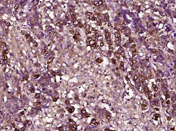
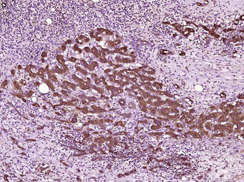
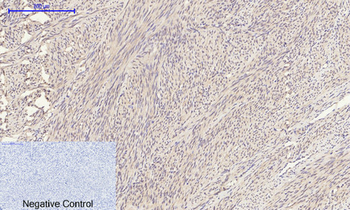
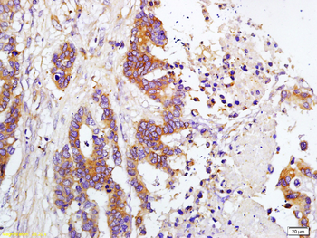
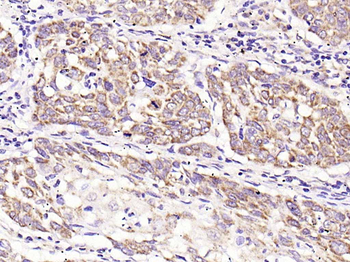
IHC-P of human colon carcinoma tissue using TIMP1 antibody (dilution at 1:250)
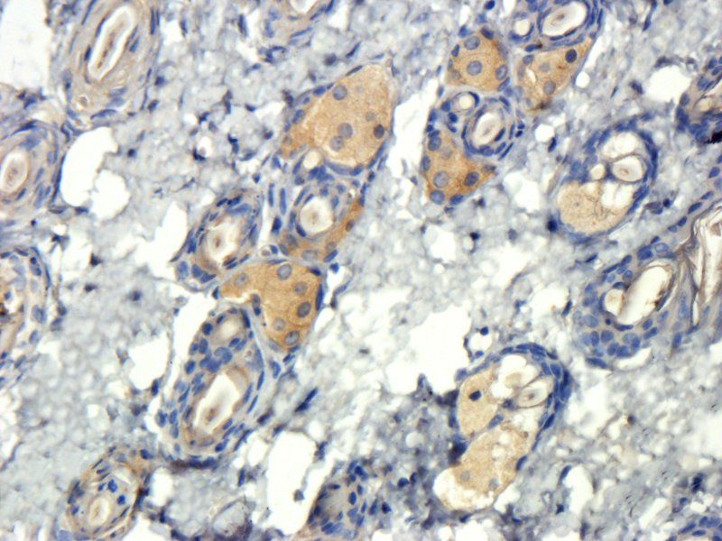
IHC-P of human colon carcinoma tissue using TIMP1 antibody (dilution at 1:250)
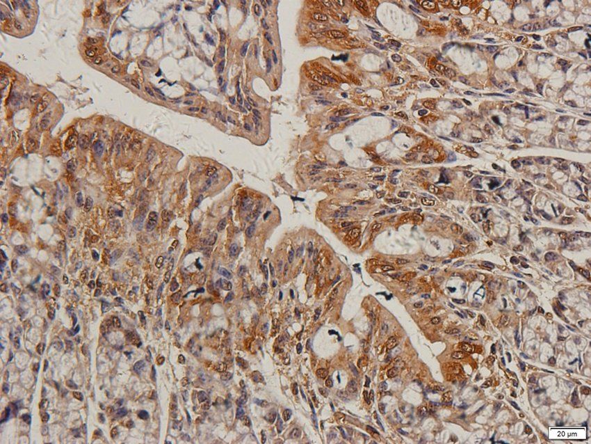
IHC-P of human colon carcinoma tissue using TIMP1 antibody (dilution at 1:250)
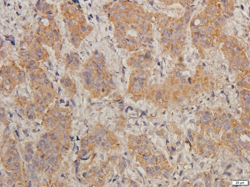
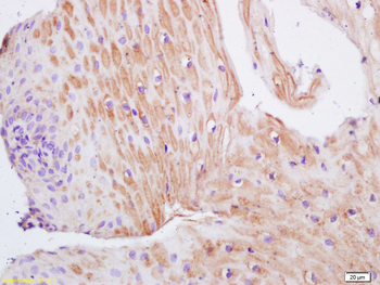
Immunohistochemical staining of mouse skin tissue using TIMP1 antibody (dilution of primary antibody - 1:400)
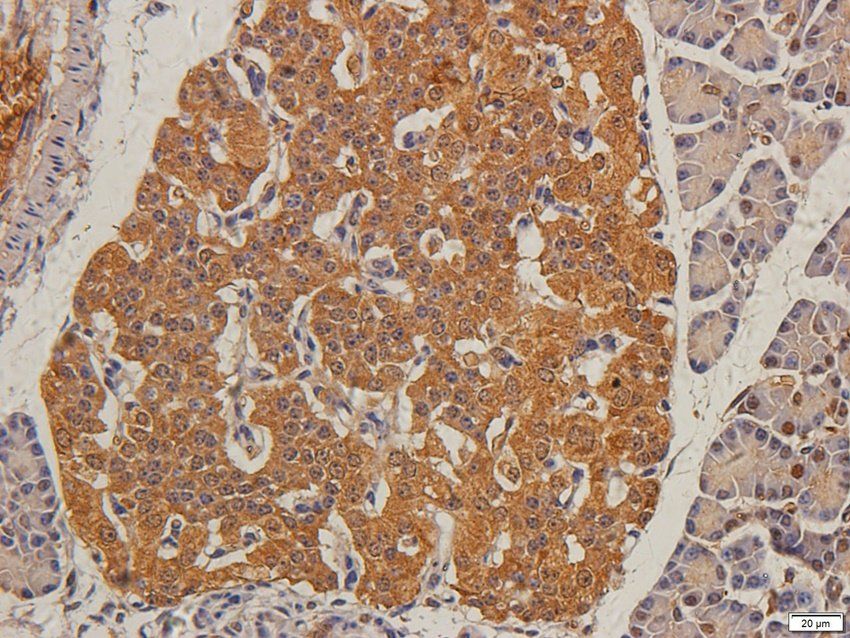
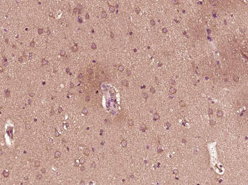
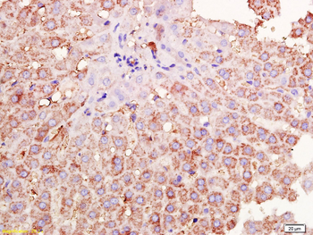
Immunohistochemical staining of paraffin embedded guinea pig colon tissue using TIMP1 antibody (2.5ug/ml)
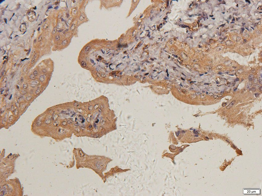
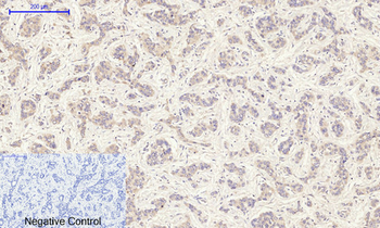
IHC-P image of human breast cancer tissue using TIMP1 antibody (2.5ug/ml)
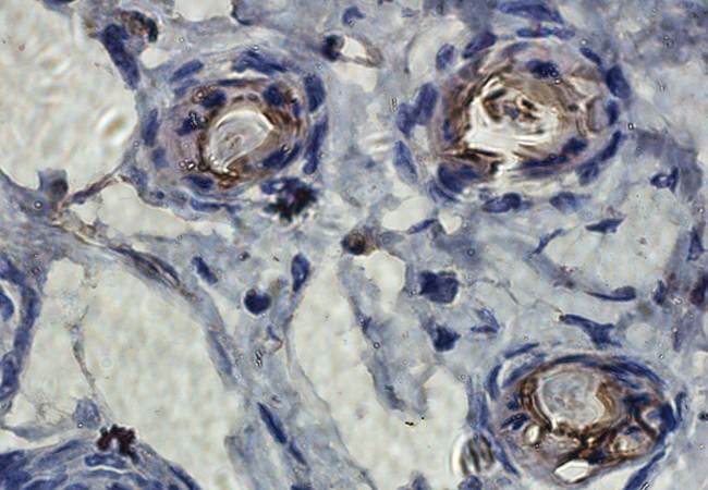
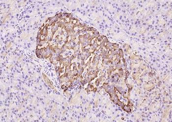
IHC-P staining of guinea pig pancreas tissue using TIMP1 antibody (2.5ug/ml)
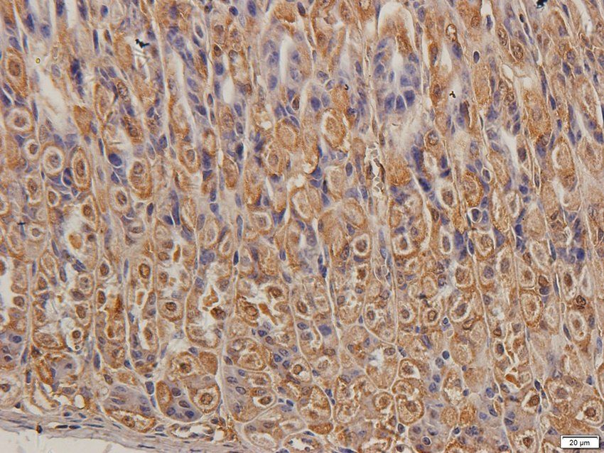
Immunohistochemical staining of rat prostate tissue using TIMP1 antibody (2.5ug/ml)
TIMP-1 Mouse Monoclonal Antibody [orb500828]
IF, IHC-Fr, IHC-P, WB
Mouse, Rat
Human, Mouse, Rat
Mouse
Monoclonal
Unconjugated
100 μl, 50 μl, 200 μlTIMP-1 Polyclonal Antibody [orb1411955]
IF, IHC-P, WB
Human, Mouse, Rat
Rabbit
Polyclonal
Unconjugated
100 μlTIMP-1 Rabbit Polyclonal Antibody [orb313247]
ELISA, IF, IHC-Fr, IHC-P, WB
Bovine, Canine, Porcine, Rat, Sheep
Human, Mouse, Rabbit
Rabbit
Polyclonal
Unconjugated
100 μl, 200 μl, 50 μlTIMP-1 Rabbit Polyclonal Antibody [orb100174]
ELISA, IF, IHC-Fr, IHC-P, WB
Bovine, Canine, Mouse, Porcine, Rabbit, Sheep
Human, Rat
Rabbit
Polyclonal
Unconjugated
100 μl, 200 μl, 50 μl


