You have no items in your shopping cart.
Cart summary
Item 1 of 3
Item 1 of 3
TIM3 antibody
Catalog Number: orb2276373

| Catalog Number | orb2276373 |
|---|---|
| Category | Antibodies |
| Description | TIM3 antibody validated for immunohistochemistry on 76 different Normal Tissues |
| Species/Host | Rabbit |
| Clonality | Monoclonal |
| Clone Number | Biorbyt-366R |
| Tested applications | IHC |
| Reactivity | Human |
| Isotype | IgG |
| Dilution range | 1:100-1:200 |
| Conjugation | Unconjugated |
| UniProt ID | Q8TDQ0 |
| Storage | Antibody with azide – store at 2 to 8°C. Antibody without azide – store at -20 to -80°C. Antibody is stable for 24 months. Non- hazardous. No MSD required. |
| Alternative names | CD366; HAVR2; Hepatitis A virus cellular receptor Read more... |
| Note | For research use only |
| Application notes | Positive Control: Tonsil: A moderate to strong TIM-3 immunostaining should be seen in a significant fraction of T-cells, while only few TIM-3 positive cells are seen in the germinal centre and the mantle zone. Negative Control: Tonsil: Epithelial cells should be negative. Cellular Localization: Cell Surface Protocol Recommendations: Manual Protocol: Freshly cut sections should be used (less than 10 days between cutting and staining). Heat-induced antigen retrieval for 5 minutes in an autoclave at 121°C in pH 7,8 Target Retrieval Solution buffer. Apply the antibody at a dilution of 1:150 at 37°C for 60 minutes. Visualization of bound antibody by the EnVision Kit (Dako, Agilent) according to the manufacturer’s directions. |
| Expiration Date | 12 months from date of receipt. |
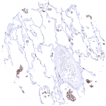
Alveolar macrophages show strong TIM 3 immunostaining.

Colon descendens, mucosa: In the colon mucosa, variable levels of TIM-3 immunostaining are seen in T-cells and in macrophages. Epithelial staining does not occur.
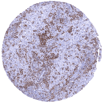
Uterus cervix TIM3 negative squamous cell carcinoma showing a large number of TIM3 positive macrophages and also lymphocytes.
- Item 1 of 6
- Item 1 of 6
- Item 1 of 5
- Item 1 of 5
- Item 1 of 5


Submit a review
Filter by Rating

- 5 stars

- 4 stars
- 3 stars
- 2 stars
- 1 stars
