You have no items in your shopping cart.
Cart summary
Item 1 of 6
Item 1 of 6
TGFB2 Antibody
Catalog Number: orb1939376

| Catalog Number | orb1939376 |
|---|---|
| Category | Antibodies |
| Description | Mouse Monoclonal Antibody (Mab) |
| Species/Host | Mouse |
| Clonality | Monoclonal |
| Clone Number | 220ct16.4.3.1 |
| Tested applications | IF, IHC-P, WB |
| Reactivity | Human |
| Isotype | IgG1,K |
| Dilution range | IF: 1:10~50, WB: 1:100~1000, WB: 1:500-1:1000, IHC-P: 1:10~50, IHC-P: 1:10~50, IHC: 1:50 |
| Form/Appearance | Purified monoclonal antibody supplied in PBS with 0.09% (W/V) sodium azide. This antibody is purified through a protein G column, followed by dialysis against PBS. |
| Conjugation | Unconjugated |
| MW | 47748 Da |
| Target | This TGFB2 monoclonal antibody is generated from mouse immunized with TGFB2 recombinant protein. |
| UniProt ID | P61812 |
| NCBI | NP_001129071.1, NP_003229.1 |
| Storage | Maintain refrigerated at 2-8°C for up to 2 weeks. For long term storage store at -20°C in small aliquots to prevent freeze-thaw cycles |
| Alternative names | Transforming growth factor beta-2, TGF-beta-2, BSC Read more... |
| Note | For research use only |
| Expiration Date | 12 months from date of receipt. |

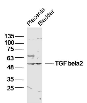
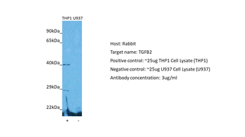
All lanes: Anti- at 1:500-1:1000 dilution. Lane 1: 293 whole cell lysate. Lane 2: K562 whole cell lysate.Lysates/proteins at 20 µg per lane. Secondary Goat Anti-mouse IgG, (H+L), Peroxidase conjugated at 1/10000 dilution. Predicted band size: 48 kDa. Blocking/Dilution buffer: 5% NFDM/TBST.
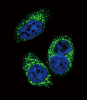
Confocal immunofluorescent analysis of TGFB2 Antibody with A549 cell followed by Alexa Fluor 488-conjugated goat anti-mouse lgG (green). DAPI was used to stain the cell nuclear (blue).

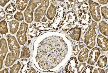
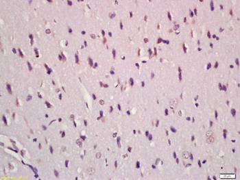
Immunohistochemical analysis of paraffin-embedded Human kidney section using Pink1. diluted at 1:50 dilution. A undiluted biotinylated goat polyvalent antibody was used as the secondary, followed by DAB staining.

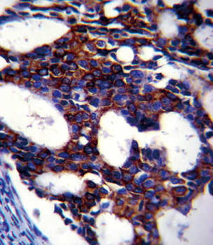
TGFB2 Antibody immunohistochemistry analysis in formalin fixed and paraffin embedded human breast carcinoma followed by peroxidase conjugation of the secondary antibody and DAB staining. This data demonstrates the use of TGFB2 Antibody for immunohistochemistry. Clinical relevance has not been evaluated.
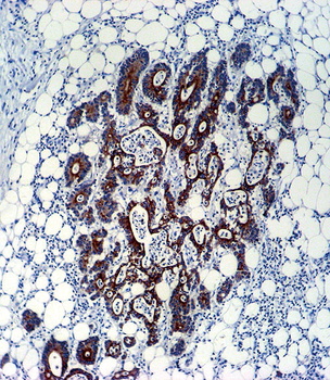
TGFB2 Antibody immunohistochemistry analysis in formalin fixed and paraffin embedded human colon carcinoma followed by peroxidase conjugation of the secondary antibody and DAB staining. This data demonstrates the use of TGFB2 Antibody for immunohistochemistry. Clinical relevance has not been evaluated.
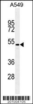
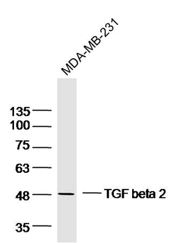
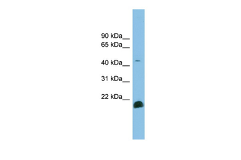
TGFB2/MB10181 antibody western blot analysis in A549 cell line lysates (35 μg/lane). This demonstrates the TGFB2/MB10181 antibody detected the TGFB2/MB10181 protein (arrow).
- Item 1 of 6
- Item 1 of 4
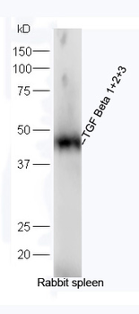
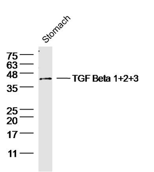
TGF Beta 1+2+3 Rabbit Polyclonal Antibody [orb7086]
FC, IF, IHC-Fr, IHC-P, WB
Bovine, Canine, Guinea pig, Porcine, Sheep
Human, Mouse, Rabbit, Rat
Rabbit
Polyclonal
Unconjugated
100 μl, 200 μl, 50 μl - Item 1 of 3
TGF beta 2 Rabbit Polyclonal Antibody [orb500662]
WB
Mouse, Rat
Human, Mouse, Rat
Rabbit
Polyclonal
Unconjugated
50 μl, 100 μl, 200 μl - Item 1 of 5
TGFB2 Rabbit Polyclonal Antibody [orb579467]
WB
Bovine, Canine, Equine, Guinea pig, Porcine, Rabbit, Rat, Sheep, Zebrafish
Human, Mouse
Rabbit
Polyclonal
Unconjugated
100 μl - Item 1 of 2
TGFB2/TGF Beta2 Antibody [orb1537317]
ELISA, IHC-P, WB
Human, Mouse
Rabbit
Polyclonal
Unconjugated
50 μg