You have no items in your shopping cart.
Cart summary
Item 1 of 4
Item 1 of 4
SUMO4 Antibody (M55 Wild type)
Catalog Number: orb1937298

| Catalog Number | orb1937298 |
|---|---|
| Category | Antibodies |
| Description | Purified Rabbit Polyclonal Antibody (Pab) |
| Species/Host | Rabbit |
| Clonality | Polyclonal |
| Clone Number | RB7262 |
| Tested applications | IHC-P, WB |
| Reactivity | Human |
| Isotype | Rabbit IgG |
| Dilution range | WB: 1:2000, WB: 1:1000, IHC-P: 1:50~100, IHC-P: 1:50~100 |
| Form/Appearance | Purified polyclonal antibody supplied in PBS with 0.09% (W/V) sodium azide. This antibody is prepared by Saturated Ammonium Sulfate (SAS) precipitation followed by dialysis against PBS. |
| Conjugation | Unconjugated |
| MW | 10653 Da |
| Target | This SUMO4 antibody is generated from rabbits immunized with a KLH conjugated synthetic peptide between 34-63 amino acids from human SUMO4. |
| UniProt ID | Q6EEV6 |
| NCBI | NP_001002255.1 |
| Storage | Maintain refrigerated at 2-8°C for up to 2 weeks. For long term storage store at -20°C in small aliquots to prevent freeze-thaw cycles |
| Alternative names | Small ubiquitin-related modifier 4, SUMO-4, Small Read more... |
| Note | For research use only |
| Expiration Date | 12 months from date of receipt. |

All lanes: Anti-SUMO4 Antibody (M55 Wild type) at 1:2000 dilution. Lane 1: 293T-17 whole cell lysate. Lane 2: 293 whole cell lysate. Lane 3: Jurkat whole cell lysate. Lane 4: Hela whole cell lysate. Lysates/proteins at 20 µg per lane. Secondary Goat Anti-Rabbit IgG, (H+L), Peroxidase conjugated at 1/10000 dilution. Predicted band size: 17 kDa. Blocking/Dilution buffer: 5% NFDM/TBST.
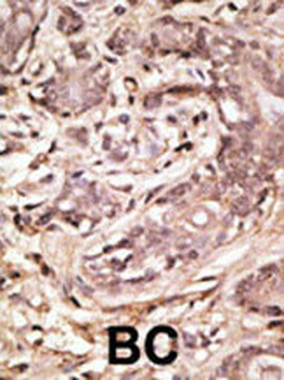
Formalin-fixed and paraffin-embedded human cancer tissue reacted with the primary antibody, which was peroxidase-conjugated to the secondary antibody, followed by AEC staining. This data demonstrates the use of this antibody for immunohistochemistry; clinical relevance has not been evaluated. BC = breast carcinoma; HC = hepatocarcinoma.
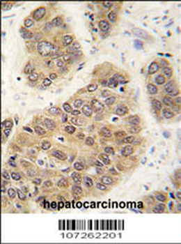
Formalin-fixed and paraffin-embedded human hepatocarcinoma tissue reacted with SUMO4 antibody (M55 Wild type), which was peroxidase-conjugated to the secondary antibody, followed by DAB staining. This data demonstrates the use of this antibody for immunohistochemistry; clinical relevance has not been evaluated.
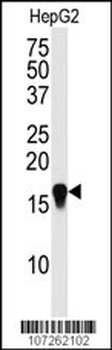
Western blot analysis of SUMO4 Antibody (M55 Wild type) in HepG2 cell line lysate (35 ug/lane). SUMO4 wild type (arrow) was detected using the purified Pab.
