You have no items in your shopping cart.
Cart summary
Item 1 of 3
Item 1 of 3
SPN Antibody
Catalog Number: orb1252702

| Catalog Number | orb1252702 |
|---|---|
| Category | Antibodies |
| Description | SPN Antibody |
| Species/Host | Mouse |
| Clonality | Monoclonal |
| Clone Number | DF-T1 |
| Tested applications | FC, IF, IHC, WB |
| Reactivity | Human |
| Isotype | IgG1, kappa |
| Immunogen | Myeloblastic KG1 cells were used as the immunogen. |
| Concentration | 0.2 mg/mL |
| Dilution range | Western Blot: 0.5-1 ug/mlFlow Cytometry: 0.5-1 ug/million cellsIF: 1-2 ug/mlIHC (FFPE): 0.5-1 ug/ml for 30 minutes at RT (1)Prediluted format : incubate for 30 min at RT (2)The concentration stated for each application is a general starting point. Variations in protocols, secondaries and substrates may require the antibody to be titered up or down for optimal performance.1. Staining of formalin-fixed tissues requires boiling tissue sections in 10mM citrate buffer, pH 6.0, for 10-20 min followed by cooling at RT for 20 minutes.2. The prediluted format is supplied in a dropper bottle and is optimized for use in IHC. After epitope retrieval step (if required), drip mAb solution onto the tissue section and incubate at RT for 30 min. |
| Form/Appearance | Liquid |
| Conjugation | Unconjugated |
| Target | SPN |
| UniProt ID | P16150 |
| Storage | Aliquot and Store at 2-8°C. Avoid freez-thaw cycles. |
| Buffer/Preservatives | PBS with 0.1 mg/ml rAlbumin and 0.05% sodium azide |
| Alternative names | SPN, GPL115, Leukosialin, Leukocyte sialoglycoprot Read more... |
| Note | For research use only |
| Application notes | Western Blot: 0.5-1 ug/mlFlow Cytometry: 0.5-1 ug/million cellsIF: 1-2 ug/mlIHC (FFPE): 0.5-1 ug/ml for 30 minutes at RT (1)Prediluted format : incubate for 30 min at RT (2)The concentration stated for each application is a general starting point. Variations in protocols, secondaries and substrates may require the antibody to be titered up or down for optimal performance.1. Staining of formalin-fixed tissues requires boiling tissue sections in 10mM citrate buffer, pH 6.0, for 10-20 min followed by cooling at RT for 20 minutes.2. The prediluted format is supplied in a dropper bottle and is optimized for use in IHC. After epitope retrieval step (if required), drip mAb solution onto the tissue section and incubate at RT for 30 min. |
| Expiration Date | 12 months from date of receipt. |
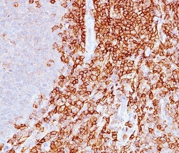
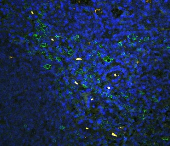
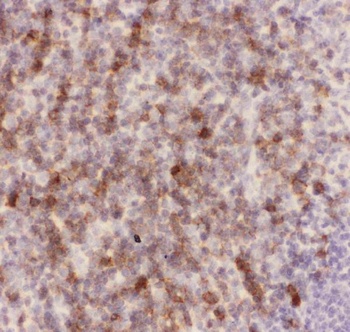
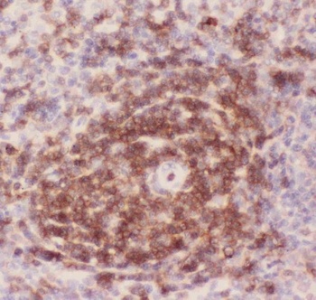
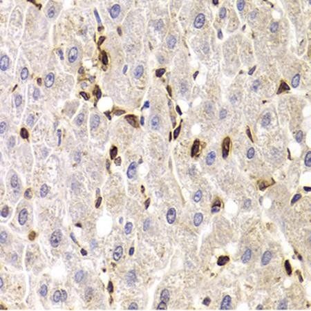
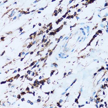
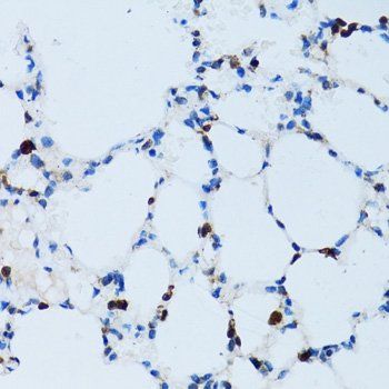
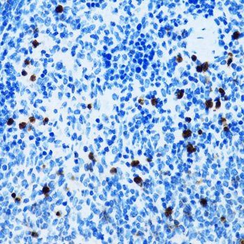
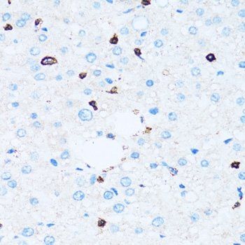
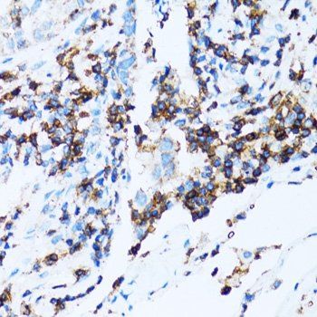
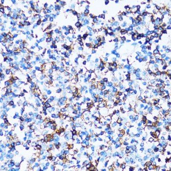
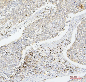
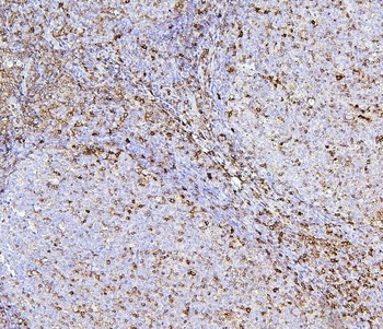
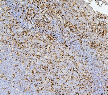
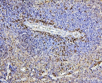
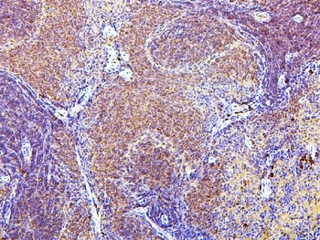
IHC staining of spleen with CD43 antibody (DF-T1).
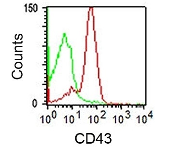
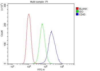
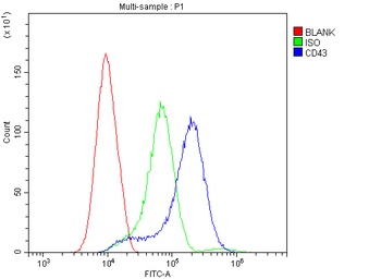
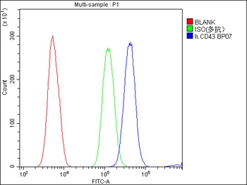
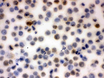
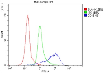
FACS staining of human lymphocytes using DF-T1 mAb (red) and isotype control antibody.
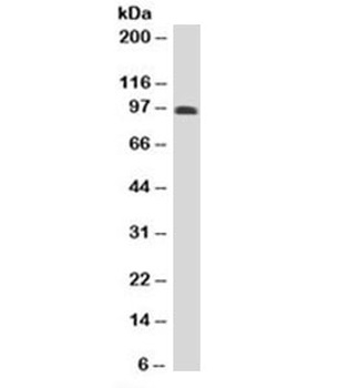
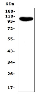
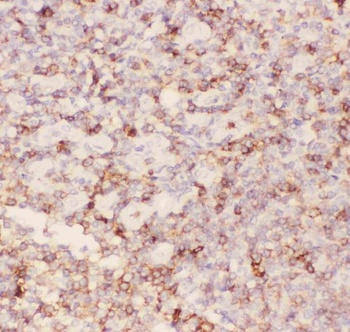
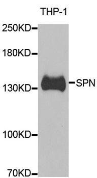
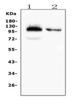
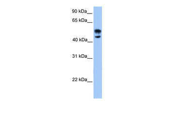
Western blot testing of human spleen lysate with CD43 antibody (clone DF-T1). Predicted molecular weight 45-115 kDa depending on glycosylation level.
- Item 1 of 9
CD43/SPN Antibody [orb196265]
FC, ICC, IF, IHC, IHC-Fr, WB
Human, Mouse, Rat
Rabbit
Polyclonal
Unconjugated
10 μg, 100 μg - Item 1 of 8
SPN antibody [orb247420]
ICC, IF, IHC, WB
Human, Mouse, Rat
Polyclonal
Unconjugated
200 μl, 100 μl, 50 μl - Item 1 of 7
CD43/SPN Antibody (monoclonal, 4I3) [orb570327]
FC, IHC, WB
Human, Mouse, Rat
Mouse
Monoclonal
Unconjugated
10 μg, 100 μg - Item 1 of 4
- Item 1 of 4
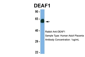
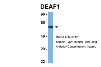
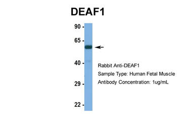
DEAF1 antibody [orb577132]
WB
Bovine, Canine, Guinea pig, Mouse, Rat, Yeast
Human
Rabbit
Polyclonal
Unconjugated
100 μl


Submit a review
Filter by Rating

- 5 stars

- 4 stars
- 3 stars
- 2 stars
- 1 stars
