You have no items in your shopping cart.
Cart summary
Item 1 of 5
Item 1 of 5
SOCS1 Antibody (N-term)
Catalog Number: orb1928279

| Catalog Number | orb1928279 |
|---|---|
| Category | Antibodies |
| Description | Purified Rabbit Polyclonal Antibody (Pab) |
| Species/Host | Rabbit |
| Clonality | Polyclonal |
| Clone Number | RB17347 |
| Tested applications | FC, IF, IHC-P, WB |
| Reactivity | Mouse, Rat |
| Isotype | Rabbit IgG |
| Antibody Type | Primary Antibody |
| Dilution range | IF: 1:10~50, WB: 1:2000, WB: 1:1000, IHC-P: 1:10~50, FC: 1:10~50 |
| Form/Appearance | Purified polyclonal antibody supplied in PBS with 0.09% (W/V) sodium azide. This antibody is prepared by Saturated Ammonium Sulfate (SAS) precipitation followed by dialysis against PBS. |
| Conjugation | Unconjugated |
| MW | 23551 Da |
| Target | This SOCS1 antibody is generated from rabbits immunized with a KLH conjugated synthetic peptide between 35-66 amino acids from the N-terminal region of human SOCS1. |
| UniProt ID | O15524 |
| NCBI | NP_003736.1 |
| Storage | Maintain refrigerated at 2-8°C for up to 2 weeks. For long term storage store at -20°C in small aliquots to prevent freeze-thaw cycles |
| Alternative names | Suppressor of cytokine signaling 1, SOCS-1, JAK-bi Read more... |
| Note | For research use only |
| Expiration Date | 12 months from date of receipt. |

Western blot analysis of SOCS1 Antibody (N-term) in mouse kidney tissue lysates (35 ug/lane). SOCS1 (arrow) was detected using the purified Pab.
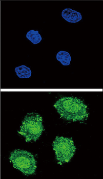
Confocal immunofluorescent analysis of SOCS1 Antibody (N-term) with 293 cell followed by Alexa Fluor 488-conjugated goat anti-rabbit lgG (green). DAPI was used to stain the cell nuclear (blue).
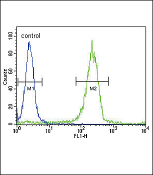
SOCS1 Antibody (N-term) flow cytometric analysis of WiDr cells (right histogram) compared to a negative control cell (left histogram). FITC-conjugated goat-anti-rabbit secondary antibodies were used for the analysis.
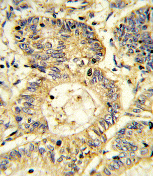
Formalin-fixed and paraffin-embedded human colon carcinoma reacted with SOCS1 Antibody (N-term), which was peroxidase-conjugated to the secondary antibody, followed by DAB staining. This data demonstrates the use of this antibody for immunohistochemistry; clinical relevance has not been evaluated.
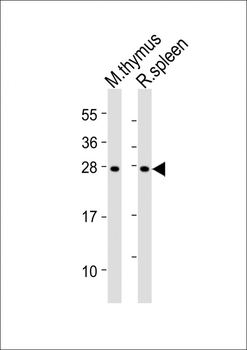
All lanes: Anti-SOCS1 Antibody (N-term) at 1:2000 dilution. Lane 1: mouse thymus lysates. Lane 2: rat spleen lysates. Lysates/proteins at 20 µg per lane. Secondary Goat Anti-Rabbit IgG, (H+L), Peroxidase conjugated at 1/10000 dilution. Predicted band size: 24 kDa. Blocking/Dilution buffer: 5% NFDM/TBST.

SOCS1 Antibody (N-term) [orb1166184]
FC, IF, IHC-P, WB
Mouse, Rat
Rabbit
Polyclonal
Unconjugated
100 μl, 30 μl
