You have no items in your shopping cart.
Cart summary
Item 1 of 7
Item 1 of 7
SMAD3 (phospho-S423) antibody
Catalog Number: orb345537

| Catalog Number | orb345537 |
|---|---|
| Category | Antibodies |
| Description | SMAD3 (phospho-S423) antibody |
| Species/Host | Rabbit |
| Clonality | Polyclonal |
| Tested applications | ELISA, FC, IHC, WB |
| Reactivity | Human |
| Isotype | IgG |
| Immunogen | Anti-SMAD3 pS423pS425 antibody was prepared from whole rabbit serum produced by repeated immunizations with a dual phosphorylated synthetic peptide corresponding to a c-terminal region with Serine 423 and Serine 425 of human SMAD3 protein. |
| Concentration | 1.16 mg/mL |
| Dilution range | ELISA: 1:75,000 - 1:300,000, FC: User Optimized, IHC: 1:500, WB: 1:2,000 - 1:20,000 |
| Form/Appearance | Liquid (sterile filtered) |
| Purity | This affinity-purified antibody is directed against the phosphorylated form of human Smad3 protein at the pS423 and pS425 residues. The product was affinity purified from monospecific antiserum by immunoaffinity purification. Antiserum was first purified against the phosphorylated form of the immunizing peptide. The resultant affinity purified antibody was then cross adsorbed against the non-phosphorylated form of the immunizing peptide. Reactivity occurs against human Smad3 pS423 and pS425 protein and the antibody is specific for the phosphorylated form of the protein. Reactivity with non-phosphorylated human Smad3 is minimal by ELISA and western blot. Expect reactivity against phosphorylated Smad1 and Smad5. Negligible reactivity is seen against other phosphorylated Smad family members. A BLAST analysis was used to suggest cross reactivity with Smad3 from human, Xenopus laevis, Xenopus tropicalis, zebrafish, rat, mouse, swine, bovine and chicken based on 100% sequence homology with the immunogen. Reactivity against homologues from other sources is not known. |
| Conjugation | Unconjugated |
| UniProt ID | P84022 |
| NCBI | 5174513 |
| Storage | Store vial at -20° C or below prior to opening. This vial contains a relatively low volume of reagent (25 µL). To minimize loss of volume dilute 1:10 by adding 225 µL of the buffer stated above directly to the vial. Recap, mix thoroughly and briefly centrifuge to collect the volume at the bottom of the vial. Use this intermediate dilution when calculating final dilutions as recommended below. Store the vial at -20°C or below after dilution. Avoid cycles of freezing and thawing. |
| Buffer/Preservatives | 0.01% (w/v) Sodium Azide |
| Alternative names | rabbit anti-SMAD3 pS423pS425 antibody, SMAD-3, SMA Read more... |
| Note | For research use only |
| Application notes | This affinity purified antibody has been tested for use in ELISA, immunohistochemistry and by western blot. Specific conditions for reactivity should be optimized by the end user. Expect a band approximately 48 kDa in size corresponding to phosphorylated Smad3 protein by western blotting in the appropriate stimulated tissue or cell lysate or extract. Less than 0.2% reactivity is observed against the non-phosphorylated form of the immunizing peptide. This antibody is phospho specific for dual phosphorylated pS423 and pS425 of Smad3. Stimulation with 2 ng/ml TGF-beta for 1 hour is suggested. |
| Expiration Date | 12 months from date of receipt. |

AcSDKP suppresses TGFβ/smad signaling and EndMT through the FGFR1/FRS2 pathway. (a) HMVECs were treated with N-FGFR1 for 48 h, and the FGFR1, TGFβR1 and TGFβR2 protein levels were analyzed by western blot. (b) HMVECs were treated with TGFβ2 in the presence or absence of N-FGFR1 for 15 min with or without AcSDKP preincubation. The p-smad3 and TGFβR1 protein levels were analyzed by western blot. Densitometric analysis of the p-smad3/smad3 and TGFβR1/β-actin levels (n=3) in each group was performed. (c) HMVECs were incubated with either N-FGFR1 in the presence or absence of TGFβ2 for 48 h with or without preincubation with AcSDKP for 2 h or with N-FGFR1 in the presence or absence of TGFβ2 for 48 h with or without 24 h of incubation with FGF2 (50 ng/mL). The CD31, SM22α, FSP1 and α-SMA protein levels were analyzed by western blot. (d) HMVECs were transfected with FRS2 siRNA (100 nM) for 48 h with or without AcSDKP preincubation. The VE-cadherin, FSP1, vimentin, SM22α and p-smad3 levels were analyzed by western blot. (e) HMVECs were treated with N-FGFR1 for 48 h or 15 min in the presence or absence of N-TGFβ (1, 2, 3) (1.0 µg/ml). The CD31, VE-cadherin, SM22α, FSP1, TGFβR1, TGFβR2 and p-smad3 levels were analyzed by western blot.

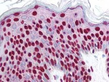
Biorbyt's affinity purified anti-Smad3 pS423 pS425 antibody was used at 2.5 ug/ml to detect signal in a variety of tissues including multi-human, multi-brain and multi-cancer slides. This image shows strong nuclear staining in the majority of epidermal keratinocytes at 40X. Tissue was formalin-fixed and paraffin embedded. The image shows localization of the antibody as the precipitated red signal, with a hematoxylin purple nuclear counterstain.

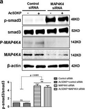
MAP4K4 deficiency induces TGFβ/smad signaling and EndMT via activation of integrin β1. (a) HMVECs were transfected with MAP4K4 siRNA (100 nM) for 48 h. Next, the cells were treated with or without AcSDKP for 2 h. The p-smad3/smad3 pathway was analyzed by western blot. Densitometric analysis of the p-smad3/smad3 levels was performed, with n=3 for each group. (b) HMVECs were treated with MAP4K4 siRNA for 48 h with or without AcSDKP treatment. The VE-cadherin, CD31, FSP1, SM22α and vimentin protein levels were analyzed by western blot. (c) HMVECs were transfected with MAP4K4 siRNA for 48 h in the presence or absence of TGFβ2 with or without AcSDKP.

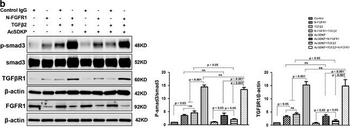
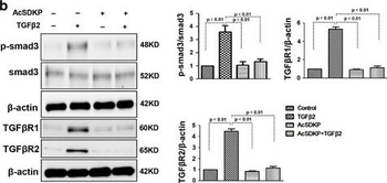
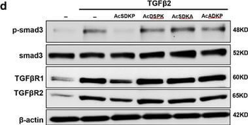
Proximity between AcSDKP and FGFR1 inhibits the TGFβ/smad signaling pathway in HMVECs. (a) HMVECs were treated with N-FGFR1 (1.5 µg/ml) for 48 h with or without preincubation with AcSDKP (100 nM) for 2 h, and the proximity between AcSDKP and FGFR1 was analyzed by the Duolink In Situ Assay. For each slide, images at a × 400 original magnification were obtained from six different areas. (b and c) HMVECs were treated with TGFβ2 (5 ng/mL) for 15 min or 48 h with or without preincubation with AcSDKP for 2 h, and the p-smad3, TGFβR1, TGFβR2 and FGFR1 levels were analyzed by western blot. Densitometric analysis of the p-smad3/smad3, TGFβR1/β-actin, TGFβR2/β-actin and FGFR1/β-actin levels from each group (n=6) were analyzed. (d and e) HMVECs were incubated with TGFβ2 for 15 min or 48 h with or without preincubation with AcSDKP or its mutants (AcDSPK, AcSDKA, AcADKP) (100 nM) for 2 h. The p-smad3/smad3, TGFβR1/β-actin, TGFβR2/β-actin and FGFR1/β-actin protein levels were analyzed by western blot .

Proximity between AcSDKP and FGFR1 inhibits the TGFβ/smad signaling pathway in HMVECs. (a) HMVECs were treated with N-FGFR1 (1.5 µg/ml) for 48 h with or without preincubation with AcSDKP (100 nM) for 2 h, and the proximity between AcSDKP and FGFR1 was analyzed by the Duolink In Situ Assay. For each slide, images at a × 400 original magnification were obtained from six different areas. (b and c) HMVECs were treated with TGFβ2 (5 ng/mL) for 15 min or 48 h with or without preincubation with AcSDKP for 2 h, and the p-smad3, TGFβR1, TGFβR2 and FGFR1 levels were analyzed by western blot. Densitometric analysis of the p-smad3/smad3, TGFβR1/β-actin, TGFβR2/β-actin and FGFR1/β-actin levels from each group (n=6) were analyzed. (d and e) HMVECs were incubated with TGFβ2 for 15 min or 48 h with or without preincubation with AcSDKP or its mutants (AcDSPK, AcSDKA, AcADKP) (100 nM) for 2 h. The p-smad3/smad3, TGFβR1/β-actin, TGFβR2/β-actin and FGFR1/β-actin protein levels were analyzed by western blot .
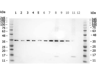
Western Blot of Rabbit anti-SMAD3 pS423 pS425 antibody. Marker: Opal Pre-stained ladder. Lane 1: HEK293 lysate (p/n orb348669). Lane 2: HeLa Lysate (p/n orb348668). Lane 3: MCF-7 Lysate (p/n orb348664). Lane 4: Jurkat Lysate. Lane 5: A431 Lysate (p/n orb348665). Lane 6: A549 Lysate (p/n orb348675). Lane 7: LNCap Lysate (p/n orb348694). Lane 8: MOLT-4 Lysate (p/n orb348696). Lane 9: Ramos Lysate. Lane 10: Raji Lysate (p/n orb348672). Lane 11: A-172 Lysate (p/n orb348708). Lane 12: NIH/3T3 Lysate (p/n orb348714). Load: 10 µg per lane. Primary antibody: SMAD3 pS423 pS425antibody at 1 ug/ml overnight at 4C. Secondary antibody: Peroxidase rabbit secondary antibody (p/n orb347654) at 1:30000 for 60 min at RT. Blocking Buffer: 1% Casein-TTBS for 30 min at RT. Predicted/Observed size: 35 kDa for SMAD3 pS423 pS425.
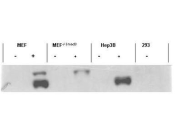
Western blot using Biorbyt's affinity purified anti-Smad3 pS423 pS425 antibody shows detection of endogenous Smad3 in stimulated cell lysates. Lysates were prepared from control cells (- lanes), or cells stimulated with 2 ng/mL TGF (+ lanes) for 1 hour. This reagent recognizes phosphorylated Smad3 and has negligible reactivity against non-phosphorylated Smad3 protein.
- Item 1 of 7
SMAD3 (phospho-S423) antibody [orb345536]
ELISA, FC, IHC, WB
Human
Rabbit
Polyclonal
Unconjugated
100 μg - Item 1 of 1
SMAD3 (phospho-S423) antibody [orb304598]
WB
Human, Mouse, Porcine, Primate, Rat, Sheep, Zebrafish
Rabbit
Polyclonal
Unconjugated
200 μl, 100 μl, 30 μl - Item 1 of 1
SMAD3 (phospho-S423/425) antibody [orb1474582]
WB
Hamster, Human, Rat
Rabbit
Monoclonal
Unconjugated
30 μl, 100 μl, 200 μl - Item 1 of 2
Smad3 (Phospho-Ser423) antibody [orb158428]
FC, ICC, WB
Porcine
Human, Mouse, Rat
Rabbit
Polyclonal
Unconjugated
100 μl, 200 μl, 50 μl 


Submit a review
Filter by Rating

- 5 stars

- 4 stars
- 3 stars
- 2 stars
- 1 stars
