You have no items in your shopping cart.
Cart summary
Item 1 of 6
Item 1 of 6
SIT antibody
Catalog Number: orb44488

| Catalog Number | orb44488 |
|---|---|
| Category | Antibodies |
| Description | Mouse Monoclonal to SIT. |
| Clonality | Monoclonal |
| Clone Number | SIT-01 |
| Tested applications | FC, IP, WB |
| Reactivity | Human |
| Isotype | Mouse IgG1 |
| Immunogen | Bacterially produced recombinant intracellular fragment of human SIT. |
| Concentration | 1 mg/ml |
| Dilution range | Flow cytometry: Recommended dilution: 1-5 μg/ml, intracellular staining. Western blotting: Recommended dilution: 1-2 μg/ml, reducing conditions. |
| Purity | Purified by protein-A affinity chromatography. |
| Conjugation | Unconjugated |
| Target | SIT |
| Entrez | 27240 |
| UniProt ID | Q9Y3P8 |
| RRID | AB_10998631 |
| Storage | Store at 2-8°C. Do not freeze. |
| Buffer/Preservatives | Phosphate buffered saline (PBS), pH 7.4, 15 mM sodium azide |
| Alternative names | SIT Read more... |
| Note | For research use only |
| Application notes | Flow cytometry: Intracellular staining. Western blotting: SIT migrates as an approximately 40 kDa protein that is reduced to approximately 20 kDa by endoglycosidase treatment. |
| Expiration Date | 12 months from date of receipt. |
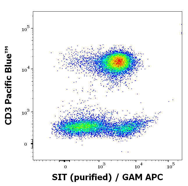
Flow cytometry multicolor intracellular staining of human peripheral whole blood stained using anti-SIT (SIT-01) purified antibody (concentration in sample 9 µg/ml, GAM APC) and anti-human CD3 (UCHT1) Pacific Blue™ antibody (20 µl reagent / 100 µl of peripheral whole blood).
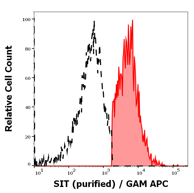
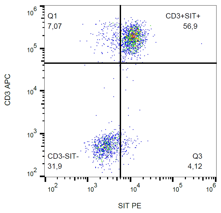
Separation of human CD3 negative SIT positive lymphocytes (red-filled) from CD3 negative SIT negative lymphocytes (black-dashed) in flow cytometry analysis (intracellular staining) of peripheral whole blood stained using anti-SIT (SIT-01) purified antibody (concentration in sample 9 µg/ml, GAM APC).
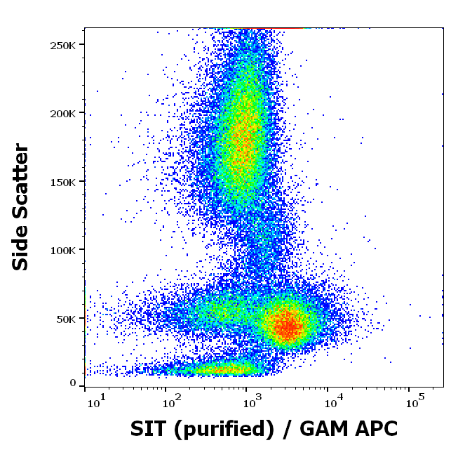
Flow cytometry intracellular staining pattern of human peripheral whole blood using anti-SIT (SIT-01) purified antibody (concentration in sample 9 µg/ml, GAM APC).
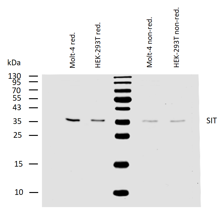
Western blotting analysis of human SIT using mouse monoclonal antibody SIT-01 on lysates of Molt-4 and HEK-293T cells under reducing and non-reducing conditions. Nitrocellulose membrane was probed with 2 µg/ml of mouse anti-SIT monoclonal antibody followed by IRDye800-conjugated anti-mouse secondary antibody. SIT was detected around 36 kDa.
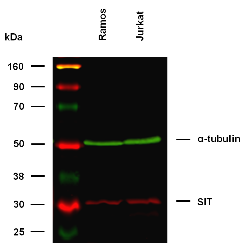
Anti-SIT Purified (clone SIT-01) works in WB application. Western blotting analysis was performed on whole cell extracts (RIPA lysis buffer) of Ramos and Jurkat cell lines, mixed and heated (100°C, 5 min) with reducing (2-mercaptoethanol) SDS-loading buffer. Samples were resolved using 10% Tris-glycine SDS gel electrophoresis. Nitrocellulose membrane blot was probed simultaneously with mouse IgG1 monoclonal antibody SIT-01 (2 µg/ml), and rat IgG2a anti-tubulin monoclonal antibody YOL1/34 (1 µg/ml) used as the loading control. Subclass-specific secondary antibodies IRDye 800CW Goat-anti-Rat IgG (green) and IRDye 680LT Goat-anti-Mouse IgG (red) were used for multiplex fluorescent Western blot detection. SIT was detected at ~32 kDa in tested cell lines.
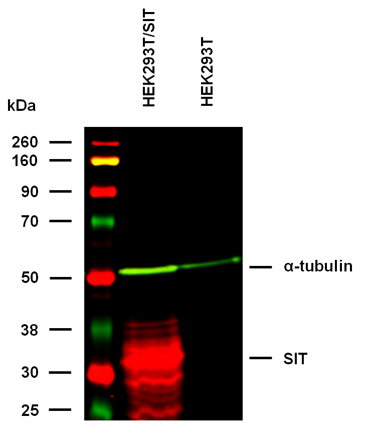
Anti-SIT Purified (clone SIT-01) specificity verification by WB. The specificity of SIT-01 antibody was assessed by comparing binding signals in HEK293T cells overexpressing the target SIT protein to wild type cells (control) with low level of endogenous protein expression. Western blotting analysis was performed on whole cell extracts (urea lysis buffer) of transfected and control cells, mixed and heated (100°C, 5 min) with reducing (2-mercaptoethanol) SDS-loading buffer. Samples were resolved using 10% Tris-glycine SDS gel electrophoresis. Nitrocellulose membrane blot was probed simultaneously with mouse IgG1 monoclonal antibody SIT-01 (2 µg/ml), and rat IgG2a anti-tubulin monoclonal antibody YOL1/34 (1 µg/ml) used as the loading control. Subclass-specific secondary antibodies IRDye 800CW Goat-anti-Rat IgG (green) and IRDye 680LT Goat-anti-Mouse IgG (red) were used for multiplex fluorescent Western blot detection.
- Item 1 of 1
- Item 1 of 1
- Item 1 of 1
SIT antibody [orb158392]
ICC, IF, IHC-Fr, IHC-P
Bovine, Human, Mouse, Porcine, Sheep
Rat
Rabbit
Polyclonal
Unconjugated
200 μl, 100 μl, 50 μl - Item 1 of 1


Submit a review
Filter by Rating

- 5 stars

- 4 stars
- 3 stars
- 2 stars
- 1 stars
