You have no items in your shopping cart.
Cart summary
Item 1 of 4
Item 1 of 4
SAE1 antibody
Catalog Number: orb18563

| Catalog Number | orb18563 |
|---|---|
| Category | Antibodies |
| Description | Goat polyclonal antibody to SAE1 |
| Species/Host | Goat |
| Clonality | Polyclonal |
| Tested applications | ELISA, FC, IF |
| Reactivity | Bovine, Canine, Human |
| Dilution range | ELISA: 1:8000, WB: 1 μg/ml |
| Conjugation | Unconjugated |
| MW | 38.4 |
| Target | SAE1 / AOS1 |
| Entrez | 10055 |
| Protein Sequence | MKGNGIVECLGPK |
| RRID | AB_10755561 |
| Storage | Aliquot and store at -20°C. Minimize freezing and thawing. |
| Buffer/Preservatives | Supplied at 0.5 mg/ml in Tris saline, 0.02% sodium azide, pH 7.3 with 0.5% bovine serum albumin. Aliquot and store at -20°C. Minimize freezing and thawing. |
| Alternative names | anti SAE1 antibody, anti AOS1 antibody, anti SUA1 Read more... |
| Note | For research use only |
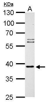
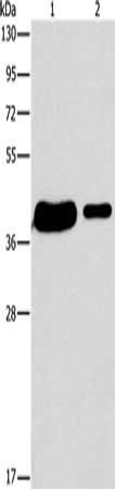
| Application notes | ELISA: Peptide ELISA: antibody detection limit dilution 1:8000.WB: Approx 40kDa band observed in Jurkat and 293 cell lysates (predicted MW of 40kDa according to NP_005491). Recommended for use at 1 μg/ml. |
| Expiration Date | 12 months from date of receipt. |

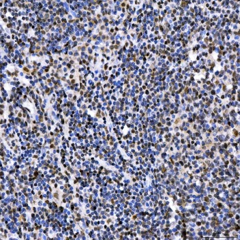
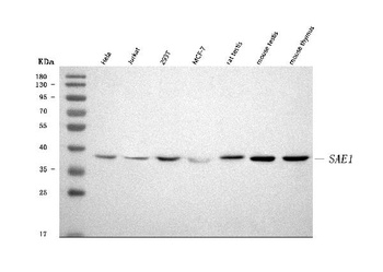
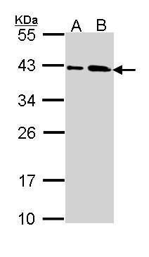
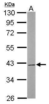
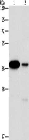
orb18563 staining (1ug/ml) of Jurkat lysate (RIPA buffer, 30ug total protein per lane). Primary incubated for 1 hour. Detected by western blot using chemiluminescence.
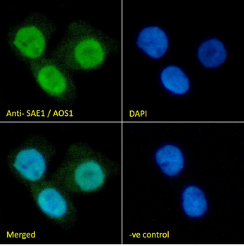
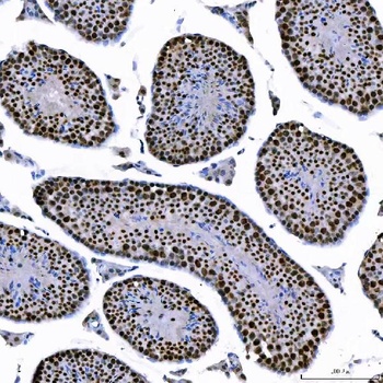
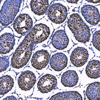
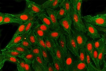
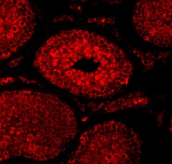
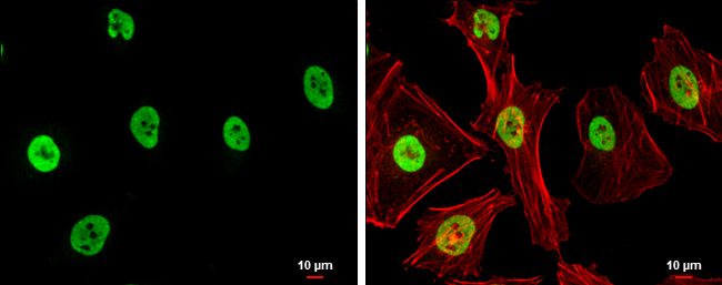
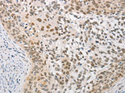
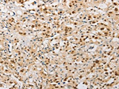
orb18563 Immunofluorescence analysis of paraformaldehyde fixed A431 cells, permeabilized with 0.15% Triton. Primary incubation 1 hr (10 µg/mL) followed by Alexa Fluor 488 secondary antibody (2 µg/mL), showing nuclear staining. The nuclear stain is DAPI (blue). Negative control: Unimmunized goat IgG (10 µg/mL) followed by Alexa Fluor 488 secondary antibody (2 µg/mL).
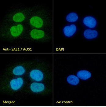
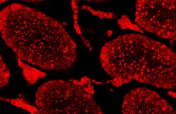
orb18563 Immunofluorescence analysis of paraformaldehyde fixed U2OS cells, permeabilized with 0.15% Triton. Primary incubation 1 hr (10 µg/mL) followed by Alexa Fluor 488 secondary antibody (2 µg/mL), showing nuclear staining. The nuclear stain is DAPI (blue). Negative control: Unimmunized goat IgG (10 µg/mL) followed by Alexa Fluor 488 secondary antibody (2 µg/mL).
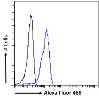
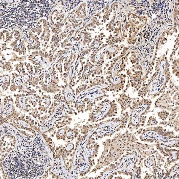
orb18563 Flow cytometric analysis of paraformaldehyde fixed A431 cells (blue line), permeabilized with 0.5% Triton. Primary incubation 1 hr (10 µg/mL) followed by Alexa Fluor 488 secondary antibody (1 µg/mL). IgG control: Unimmunized goat IgG (black line) followed by Alexa Fluor 488 secondary antibody.
- Item 1 of 12
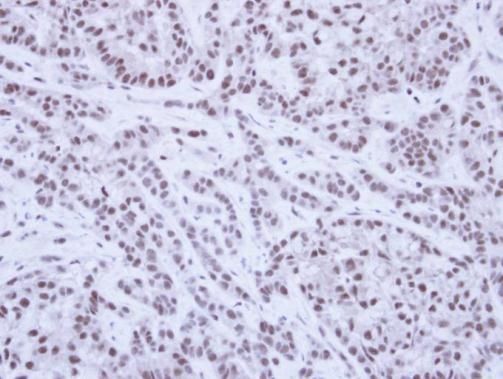
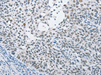
SAE1 Antibody [orb1402189]
ELISA, ICC, IF, IHC, WB
Human, Mouse, Rat
Rabbit
Polyclonal
Unconjugated
10 μg, 100 μg - Item 1 of 6
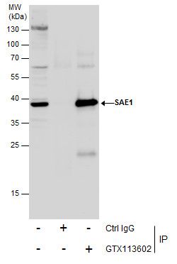
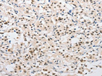
SUMO1 activating enzyme subunit 1 Antibody [orb556873]
ICC, IHC-P, IP, WB
Human, Mouse, Rat
Rabbit
Polyclonal
Unconjugated
100 μl - Item 1 of 4
- Item 1 of 3
- Item 1 of 3


Submit a review
Filter by Rating

- 5 stars

- 4 stars
- 3 stars
- 2 stars
- 1 stars
