You have no items in your shopping cart.
Cart summary
Item 1 of 1
RREB1 antibody
Catalog Number: orb344631

| Catalog Number | orb344631 |
|---|---|
| Category | Antibodies |
| Description | RREB1 antibody |
| Species/Host | Rabbit |
| Clonality | Polyclonal |
| Tested applications | ELISA, WB |
| Reactivity | Mouse |
| Isotype | IgG |
| Immunogen | Anti-RREB1 antibody was prepared from whole rabbit serum produced by repeated immunizations with a recombinant protein corresponding to mouse RREB1 protein. |
| Concentration | 1.0 mg/ml |
| Dilution range | ELISA: 1:2,000 - 1:15,000, WB: 1:500 - 1:2,000 |
| Form/Appearance | Liquid (sterile filtered) |
| Purity | Anti-RREB1 antibody is directed against the mouse RREB1 protein. The product was Protein A purified from monospecific antiserum by immunoaffinity purification. Reactivity against homologues from other sources is not known. |
| Conjugation | Unconjugated |
| UniProt ID | Q3UH06 |
| NCBI | AAX83010.1 |
| Storage | Store vial at -20° C prior to opening. Aliquot contents and freeze at -20° C or below for extended storage. Avoid cycles of freezing and thawing. Centrifuge product if not completely clear after standing at room temperature. This product is stable for several weeks at 4° C as an undiluted liquid. Dilute only prior to immediate use. |
| Buffer/Preservatives | 0.01% (w/v) Sodium Azide |
| Alternative names | rabbit anti-RREB1 Antibody, rabbit anti-RREB 1 Ant Read more... |
| Note | For research use only |
| Application notes | This Protein A purified antibody has been tested for use in ELISA and by western blot. Specific conditions for reactivity should be optimized by the end user. Expect a predominant band approximately 90-180 kDa in size corresponding to RREB1 by western blotting in the appropriate cell lysate or extract. Multiple bands seen in the above western blot may indicate cross-reactive isoforms or truncated protein products. |
| Expiration Date | 12 months from date of receipt. |
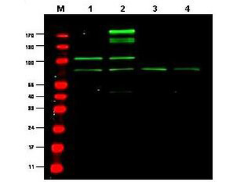
Western blot using Biorbyt's Protein A Purified anti-RREB1 antibody shows detection of a predominant band believed to be RREB1 in various cell lysates (1 - HEK293, 2 - RFP-RREB transfected HEK293, 3 - M460 and 4 - T1165). All lysates were loaded at 20 µg per lane and separated by SDS-PAGE. After transfer to nitrocellulose, the membrane was probed with the primary antibody diluted to 1:1000. The membrane was washed and reacted with IRDye800 conjugated Gt-a-Rabbit IgG [H&L] MX.
- Item 1 of 2
- Item 1 of 1
- Item 1 of 1
- Item 1 of 1
- Item 1 of 1


Submit a review
Filter by Rating

- 5 stars

- 4 stars
- 3 stars
- 2 stars
- 1 stars
