You have no items in your shopping cart.
Cart summary
Item 1 of 6
Item 1 of 6
ROR1 Antibody
Catalog Number: orb1263013

| Catalog Number | orb1263013 |
|---|---|
| Category | Antibodies |
| Description | ROR1 Antibody |
| Species/Host | Rabbit |
| Clonality | Polyclonal |
| Tested applications | FC, IHC-P, WB |
| Reactivity | Human, Mouse |
| Isotype | Rabbit Ig |
| Immunogen | This ROR1 antibody is generated from rabbits immunized with recombinant human ROR1 protein (aa region: 112 - 399). |
| Concentration | batch dependent |
| Dilution range | For FACS starting dilution is: 1:25For IHC-P starting dilution is: 1:25For WB starting dilution is: 1:1000 |
| Form/Appearance | Liquid |
| Conjugation | Unconjugated |
| MW | 104 kDa |
| Target | ROR1 |
| UniProt ID | Q01973 |
| NCBI | Q01973 |
| Storage | Store at 4°C for three months and -20°C, stable for up to one year. As with all antibodies care should be taken to avoid repeated freeze thaw cycles. Antibodies should not be exposed to prolonged high temperatures. |
| Buffer/Preservatives | Supplied in PBS with 0.09% (W/V) sodium azide. |
| Alternative names | Tyrosine-protein kinase transmembrane receptor ROR Read more... |
| Note | For research use only |
| Application notes | For FACS starting dilution is: 1:25For IHC-P starting dilution is: 1:25For WB starting dilution is: 1:1000 |
| Expiration Date | 12 months from date of receipt. |

Overlay histogram showing A549 cells stained with Antibody (green line). The cells were fixed with 2% paraformaldehyde (10 min). The cells were then icubated in 2% bovine serum albumin to block non-specific protein-protein interactions followed by the antibody (1:25 dilution) for 60 min at 37°C. The secondary antibody used was Goat-Anti-Rabbit IgG, Conjugated Highly Cross-Adsorbed at 1/200 dilution for 40 min at 37°C. Isotype control antibody (blue line) was rabbit IgG (1ug/1x10^6 cells) used under the same conditions. Acquisition of > 10000 events was performed.

Antibody staining ROR1 in human kidney tissue sections by Immunohistochemistry (IHC-P - paraformaldehyde-fixed, paraffin-embedded sections).

Antibody staining ROR1 in Human heart tissue sections by Immunohistochemistry (IHC-P - paraformaldehyde-fixed, paraffin-embedded sections).

Immunohistochemical analysis of paraffin-embedded H. heart section using ROR1 Antibody. Antibody was diluted at 1:25 dilution. A undiluted biotinylated goat polyvalent antibody was used as the secondary, followed by DAB staining.

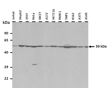
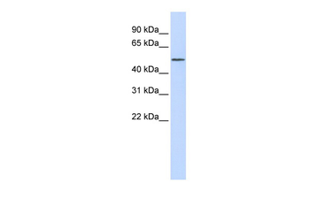
Western blot analysis of lysates from K562 cell line, human lung, mouse kidney, mouse heart tissue (from left to right), using ROR1 Antibody at 1:1000 at each lane.

Formalin-fixed and paraffin-embedded human lung carcinoma tissue reacted with the ROR1 antibody, which was peroxidase-conjugated to the secondary antibody, followed by DAB staining.
- Item 1 of 6
- Item 1 of 5
- Item 1 of 5
- Item 1 of 5
RORA antibody [orb329792]
WB
Bovine, Canine, Equine, Goat, Guinea pig, Rabbit, Rat, Zebrafish
Human, Mouse
Rabbit
Polyclonal
Unconjugated
100 μl - Item 1 of 4
Anti-ROR1 Reference Antibody [orb1806307]
ELISA, FA, FACS, Kinetics
Human
Monoclonal
Unconjugated
50 μg, 100 μg, 1 mg, 5 mg


Submit a review
Filter by Rating

- 5 stars

- 4 stars
- 3 stars
- 2 stars
- 1 stars
