You have no items in your shopping cart.
Cart summary
Item 1 of 3
Item 1 of 3
Rhodopsin Antibody: APC
Catalog Number: orb147737

| Catalog Number | orb147737 |
|---|---|
| Category | Antibodies |
| Description | Mouse monoclonal to Rhodopsin (APC). Rhodopsin consists of the protein moiety opsin and a reversibly covalently bound cofactor, retinal. Opsin, a bundle of seven membrane embedded alpha-helices, binds retinal, a photo reactive chromophore, in a central pocket. In addition to being the pigment of the retina that is responsible for both the formation of the photoreceptor cells, its function is to specifically convey information stored in the specific geometry of the chormophore to the surface of the molecule upon light absorption. In the active state, rhodopsin activates transduction, a GTP binding protein. Once activated, transduction promotes the hydrolysis of cGMP by phosphodiesterase. Rhodopsins activity is believed to be shut off by its phosphorylation followed by binding of the soluble protein arrestin. Mutations in the rhodopsin gene lead to retinitis pigmentosa, which can be inherited as an autosomal dominant, an autosomal recessive or an X-linked recessive disorder.. |
| Species/Host | Mouse |
| Clonality | Monoclonal |
| Clone Number | 4D2 |
| Tested applications | ICC, IF, IHC |
| Reactivity | Amphibian, Aves, Bovine, Fish, Human, Mouse |
| Isotype | IgG1 |
| Immunogen | Bovine Rhodopsin |
| Concentration | 1 mg/ml |
| Dilution range | WB (1:1000), IHC (1000) |
| Conjugation | APC |
| MW | 40kDa |
| Target | Rhodopsin |
| Entrez | 509933 |
| UniProt ID | P02699 |
| NCBI | NP_001014890.1 |
| Storage | Conjugated antibodies should be stored according to the product label |
| Buffer/Preservatives | 95.64mM Phosphate, 2.48mM MES and 2mM EDTA |
| Alternative names | OPN2 antibody, opsd antibody, opsin 2 antibody, op Read more... |
| Note | For research use only |
| Application notes | 1 µg/ml of SMC-176 was sufficient for detection of rhodopsin in 10 µg of rat eye lysate by colorimetric immunoblot analysis using Goat anti-mouse IgG:HRP as the secondary antibody. |
| Expiration Date | 12 months from date of receipt. |

Immunohistochemistry analysis using Mouse Anti-Rhodopsin Monoclonal Antibody, Clone 4D2. Tissue: retina. Species: Mouse. Primary Antibody: Mouse Anti-Rhodopsin Monoclonal Antibody at 1:1000. Secondary Antibody: FITC Goat Anti-Mouse (green). Counterstain: DAPI (blue) nuclear stain. Localization: Staining of photoreceptor outer segment (OS). Other layers of the retina: IS – inner segment; ONL – outer nuclear layer; OPL – outer plexiform layer; INL – inner nuclear layer; IPL – inner plexiform layer; GCL – ganglion cell layer.
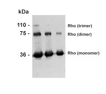
Western Blot analysis of Bovine photoreceptor membranes showing detection of Rhodopsin protein using Mouse Anti-Rhodopsin Monoclonal Antibody, Clone 4D2. Lane 1: MW ladder. Lane 2: 10 ug. Lane 3: 5 ug. Lane 4: 2.5 ug. Primary Antibody: Mouse Anti-Rhodopsin Monoclonal Antibody at 1:1000.
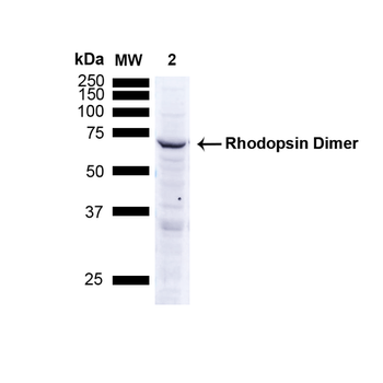
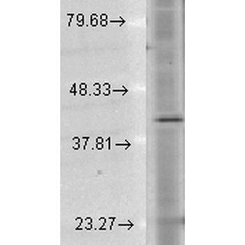
Western Blot analysis of Human A549 cells showing detection of ~38.9kDa Rhodopsin protein using Mouse Anti-Rhodopsin Monoclonal Antibody, Clone 4D2. Lane 1: MW ladder. Lane 2: Human A549 Cells 15 ug). Load: 15 ug. Block: 5% Skim Milk Powder in TBST. Primary Antibody: Mouse Anti-Rhodopsin Monoclonal Antibody at 1:1000 for 2.5 hours at RT with shaking. Secondary Antibody: Goat anti-mouse IgG:HRP at 1:1000 for 1 hour at RT with shaking. Color Development: Chemiluminescent for HRP (Moss) for 5 min in RT. Predicted/Observed Size: ~38.9kDa. Other Band (s): Band appears at ~75 kDa indicating detection of the Rhodopsin dimer.
- Item 1 of 2

PDE6D Rabbit Polyclonal Antibody (APC) [orb1001270]
ICC, IF
Bovine, Equine, Gallus, Human, Mouse, Rabbit, Zebrafish
Rat
Rabbit
Polyclonal
APC
100 μlRhodopsin/RP4/RHO Rabbit Polyclonal Antibody (APC) [orb1001414]
ICC, IF
Bovine, Canine, Human, Mouse, Rabbit, Rat
Rabbit
Polyclonal
APC
100 μlGRK1 Rabbit Polyclonal Antibody (APC) [orb1005590]
IF
Bovine, Gallus
Human, Mouse, Rat
Rabbit
Polyclonal
APC
100 μlGRK1 Rabbit Polyclonal Antibody (APC-Cy7) [orb2549519]
IF
Bovine, Gallus
Human, Mouse, Rat
Rabbit
Polyclonal
APC/Cy7
100 μl