You have no items in your shopping cart.
Cart summary
Item 1 of 2
Item 1 of 2
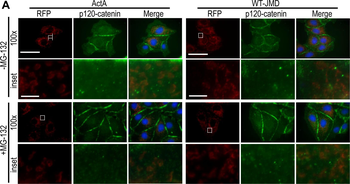
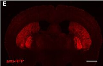
RFP antibody
Catalog Number: orb345939

| Catalog Number | orb345939 |
|---|---|
| Category | Assays and Kits |
| Description | RFP antibody |
| Clonality | Polyclonal |
| Tested applications | WB |
| Reactivity | Other |
| Dilution range | WB: User Optimized |
| Form/Appearance | n/a |
| Purity | Biorbyt’ Chemiluminescent Western Blot Kit for RFP combines all of the necessary reagents with a rapid proven protocol and the extremely high signal detection of our luminol chemiluminescent substrate for the detection of recombinant proteins containing wild type Red Fluorescent Protein (DsRed or RFP) and its variants such as mBanana, mCherry, DsRed2, E2-Crimson, mOrange, mOrange2, mPlum, mRaspberry, mStrawberry and tdTomato. The Chemiluminescent Western Blot Kit design includes straightforward procedures and color-coding to allow for ease of use. This kit contains sufficient substrate for up to 30 mini blots at 7.5 x 8 cm2 (1,800 cm2) and is stable for at least 1 year when stored as indicated. |
| Conjugation | Unconjugated |
| UniProt ID | Q9U6Y8 |
| Storage | See kit insert for complete instructions. |
| Buffer/Preservatives | Wash buffers MUST NOT contain SODIUM AZIDE or other inhibitors of peroxidase activity! |
| Alternative names | Western Blotting Kit, Chemiluminescent Kit, Peroxi Read more... |
| Note | For research use only |
| Application notes | Use Biorbyt’ Anti-RFP Chemiluminescent Kit for Western Blotting for detection of RFP-tagged recombinant proteins by western blot. This kit is useful for both western blotting and dot blotting methods. Please read the entire product insert prior to use. |
| Expiration Date | Please enquire. |
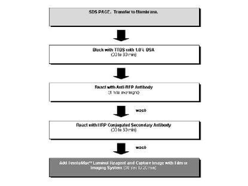
Flow Diagram for RFP Chemiluminescent Western Blot Procedure.
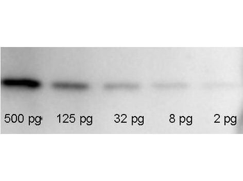
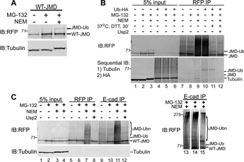
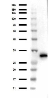
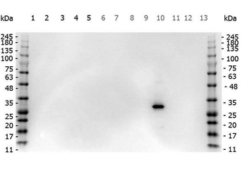
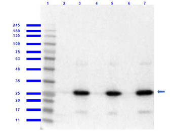
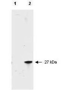
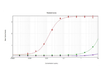
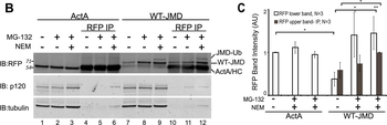
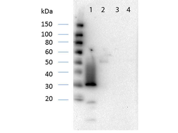
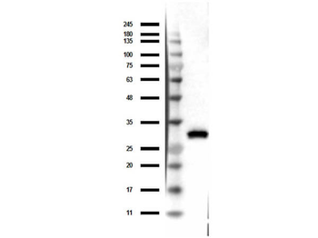
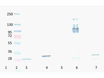
RFP Western Blot: Known amounts of recombinant RFP and GFP protein (p/n orb345957) were spiked into a HeLa cell-derived lysates (p/n orb348668) and separated by SDS-PAGE using a 4-20% gradient gel. Proteins were transferred onto a nitrocellulose membrane for 1 h at 100 mV. The membrane was blocked with TTBS (p/n orb348540) supplemented with 1% BSA for 1 h at 4°C prior to probing the blot with the anti-GFP monoclonal antibody (p/n orb345329) diluted 1:1000 for 40 min. Detection of the primary antibody by the HRP-conjugated anti-Mouse IgG (p/n orb347544) was performed at a dilution of 1:20000 for 1 h at 4°C. FemtoMax Super Sensitive Chemiluminescent Luminol Substrate was used for signal detection.
- Item 1 of 45
- Item 1 of 45
- Item 1 of 2
- Item 1 of 12
- Item 1 of 12


Submit a review
Filter by Rating

- 5 stars

- 4 stars
- 3 stars
- 2 stars
- 1 stars
