You have no items in your shopping cart.
Cart summary
Item 1 of 4
Item 1 of 4
rCan f 1 (Canis familiaris 1.0101)
Catalog Number: orb1088526

| Catalog Number | orb1088526 |
|---|---|
| Category | Proteins |
| Description | Recombinant protein rCan f 1 is expressed in Escherichia coli. DNA sequence encoding 169 AAs was fused with Strep-tag at the N-terminus. A calculated molecular mass of recombinant protein is 18,8 kDa. |
| Tested applications | ELISA, FC, WB |
| Purity | Purified by sequential steps of ion exchange and affinity chromatography. Lyophilized from a 0.2 μm filtered solution in Storage buffer (100 mM Tris, 150 mM NaCl, 0.01% Tween 20, pH 8.0). |
| UniProt ID | O18873 |
| Storage | Store at -20°C to -80°C. Reconstitute in sterile deionized water. Use reconstituted product immediately or aliquot for further storage at -20°C to -80°C. |
| Note | For research use only |
| Expiration Date | 12 months from date of receipt. |
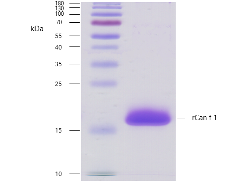
Recombinant allergen rCan f 1 purity verification. 5 µg of rCan f 1 with > 95 % purity checked by Coomassie Brilliant Blue stained SDS-PAGE.
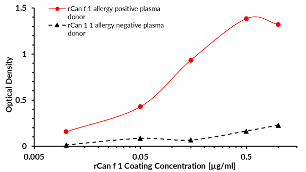
ELISA (enzyme-linked immunosorbent assay) test was designed to prove the bond between the coated target recombinant allergen rCan f 1 and allergen-specific human plasma IgG4 antibodies of Canis familiaris positive donor. A measurable signal was subsequently generated by the addition of biotin labeled anti-human IgG4 (detection) antibody, Streptavidin-HRP and substrate solution (TMB). The intensity of the signal is proportional to the amount of coated rCan f 1.
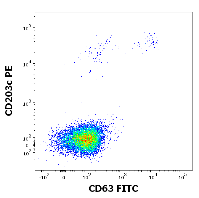
Flow cytometry dot-plot staining pattern of rCan f 1 recombinant allergen stimulated human peripheral whole blood lymphocytes and basophils of a proven allergic donor stained using anti-human CD63 (MEM-259) FITC and anti-human CD203c (NP4D6) PE antibodies of BasoFlowEx kit.
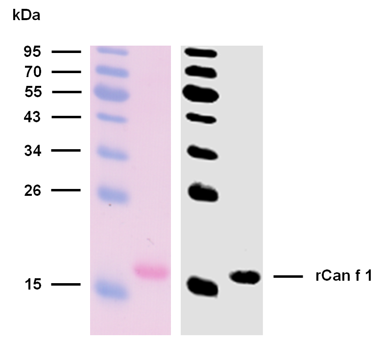
Reactivity of rCan f 1 with sIgE antibodies in plasma of a patient with confirmed presence (routine clinical test) of specific IgE antibodies to dog epithelium allergen extract. Western blotting analysis was performed on recombinant allergen rCan f 1 mixed and heated (100°C, 5 min) with reducing SDS loading buffer. Sample was resolved using 15% Tris-glycine gel electrophoresis. Nitrocellulose membrane blot was stained with Ponceau S to verify protein transfer. After thorough washing and blocking steps, the membrane was incubated with plasma of an allergic patient (1:10). Captured rCan f 1 sIgE antibodies were detected by biotinylated anti-human IgE secondary antibody (2 μg/ml) followed by IRDye 800CW Streptavidin (1:5000).
Submit a review
Filter by Rating

- 5 stars

- 4 stars
- 3 stars
- 2 stars
- 1 stars
