You have no items in your shopping cart.
Cart summary
Item 1 of 2
Item 1 of 2
RBL2 antibody
Catalog Number: orb750457

| Catalog Number | orb750457 |
|---|---|
| Category | Antibodies |
| Description | RBL2 antibody |
| Species/Host | Rabbit |
| Clonality | Polyclonal |
| Tested applications | ELISA, IHC, IP, WB |
| Reactivity | Mouse |
| Isotype | Antiserum |
| Immunogen | Rb2 (p130) peptide corresponding to a region near the C-terminus of the human protein conjugated to Keyhole Limpet Hemocyanin (KLH). |
| Concentration | 85 mg/mL |
| Dilution range | ELISA: 1:5,000 - 1:20,000, IHC: 1:200 - 1:1,000, IP: 1:100, WB: 1:500 - 1:2,000 |
| Form/Appearance | Liquid (sterile filtered) |
| Purity | This product was prepared from monospecific antiserum by delipidation and defibrination. Antiserum will specifically react with a 130 kDa Rb2 protein from human, rat and mouse tissue. No reaction was observed against other related tumor suppressor proteins. Cross reactivity with Rb2 (p130) from other species may also occur. |
| Conjugation | Unconjugated |
| UniProt ID | Q08999 |
| NCBI | 172072597 |
| Storage | Store vial at -20° C prior to opening. Aliquot contents and freeze at -20° C or below for extended storage. Avoid cycles of freezing and thawing. Centrifuge product if not completely clear after standing at room temperature. This product is stable for several weeks at 4° C as an undiluted liquid. Dilute only prior to immediate use. |
| Buffer/Preservatives | 0.01% (w/v) Sodium Azide |
| Alternative names | rabbit anti-p130 Antibody, rabbit anti-Rb2 antibod Read more... |
| Note | For research use only |
| Application notes | Anti-p130 has been tested by western blot and immunohistochemistry and is suitable for ELISA, immunoprecipitation, immunoblotting, immunohistochemistry, and other immunological methods requiring high titer and specificity. |
| Expiration Date | 12 months from date of receipt. |
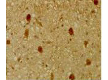
Immunohistochemical staining of mouse tissue using anti-pRb2/p130 antiserum. The staining shows the location of pRb2/p130 in developing mouse tissue. Other detection systems should yield similar results. Sections were cut at 5-7 µM, mounted on glass and dried overnight at 37°C. All sections were deparaffinized in xylene, rehydrated through a graded alcohol series and washed in phosphate-buffered saline (PBS). PBS was used for all subsequent washes and for antiserum dilution. Tissue sections were quenched sequentially in 0.5% hydrogen peroxide and blocked with diluted 10% normal goat anti-rabbit serum. Slides were incubated at 20°C for 1 h with rabbit anti-pRb2/p130 (1:500) dilution, washed, and then reacted with diluted goat anti-rabbit biotinylated antibody for 30 min. Slides were then reacted with streptavidin-peroxidase conjugate for 30 min at 20°C. Diaminobenzidine was used as the final chromogen. Negative controls for each tissue section were prepared by substituting the primary antiserum with pre-immune serum.
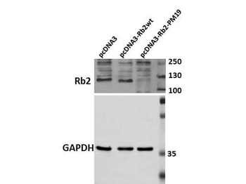
Western Blot of Rabbit Anti-Rb2 p130 Antibody. Lane 1: HEK 293 pcDNA3. Lane 2: HEK 293 pcDNA3-Rb2wt. Lane 3: HEK 293 pcDNA3-Rb2-PM19. Load: 30 µg per lane. Primary antibody: Anti-Rb2 antibody at 1:250 for overnight at 4°C. Secondary antibody: IRDye800™ rabbit secondary antibody at 1:10000 for 45 min at RT. Block: 5% BLOTTO overnight at 4°C. Predicted/Observed size: 130 kDa for p130/Rb2.
- Item 1 of 4
- Item 1 of 3
RBL2 Antibody [orb1564332]
ICC, IHC-Fr, IHC-P, WB
Human
Rabbit
Monoclonal
Unconjugated
100 μl, 50 μl, 20 μl - Item 1 of 1
- Item 1 of 2
- Item 1 of 2


Submit a review
Filter by Rating

- 5 stars

- 4 stars
- 3 stars
- 2 stars
- 1 stars
