You have no items in your shopping cart.
Cart summary
Item 1 of 6
Item 1 of 6
Rabbit anti-PD-L1 Recombinant Monoclonal Antibody
Catalog Number: orb1519952

| Catalog Number | orb1519952 |
|---|---|
| Category | Antibodies |
| Description | PD-L1 Antibody |
| Species/Host | Rabbit |
| Clonality | Recombinant |
| Clone Number | BLR020E |
| Tested applications | FC, ICC, IF, IHC, WB |
| Reactivity | Human |
| Immunogen | Between 240 and C-terminus |
| Concentration | 50 µg/ml |
| Dilution range | Flow-Cyt Fixed in 4% formaldehyde and permeabilized with 90% methanol. 2 µl per 1 x 10^6 cells. ICC-IF 1:100 - 1:500. Epitope retrieval with citrate buffer pH 6.0 is recommended for FFPE cell sections. Immunohistochemistry (IHC) 1:100 - 1:500. Epitope retrieval with citrate buffer pH 6.0 is recommended for FFPE tissue sections. IHC-IF 1:100 - 1:500. Epitope retrieval with citrate buffer pH 6.0 is recommended for FFPE tissue sections. Immunoprecipitation (IP) 20 µl/mg lysate. Multiplex Immunofluorescence (mIF) 1:250. Western Blot (WB) 1:1000 |
| Form/Appearance | Whole IgG |
| Purity | Recombinant antibody was purified from cell culture supernatant |
| Conjugation | Unconjugated |
| Target | PD-L1 |
| UniProt ID | Q9NZQ7 |
| NCBI | NP_054862.1 |
| Storage | 2 - 8°C |
| Buffer/Preservatives | Borate Buffered Saline (BBS) pH 8.2 with 0.1% rAlbumin and 0.09% Sodium Azide |
| Alternative names | PDCD1L1; Programmed death ligand 1; programmed cel Read more... |
| Background | PD-L1 is an immune inhibitory receptor ligand that is expressed by hematopoietic and non-hematopoietic cells, such as T cells and B cells and various types of tumor cells. PD-L1 is a type I transmembrane protein that has immunoglobulin V-like and C-like domains. Interaction of this ligand with its receptor inhibits T-cell activation and cytokine production. During infection or inflammation of normal tissue, this interaction is important for preventing autoimmunity by maintaining homeostasis of the immune response. In tumor microenvironments, this interaction provides an immune escape for tumor cells through cytotoxic T-cell inactivation. Expression of this gene in tumor cells is considered to be prognostic in many types of human malignancies, including colon cancer and renal cell carcinoma |
| Note | For research use only |
| Application notes | All western blot analysis is performed using 5% Milk-TBST for blocking and as antibody diluent. Primary antibody is incubated overnight. |
| Expiration Date | 12 months from date of receipt. |
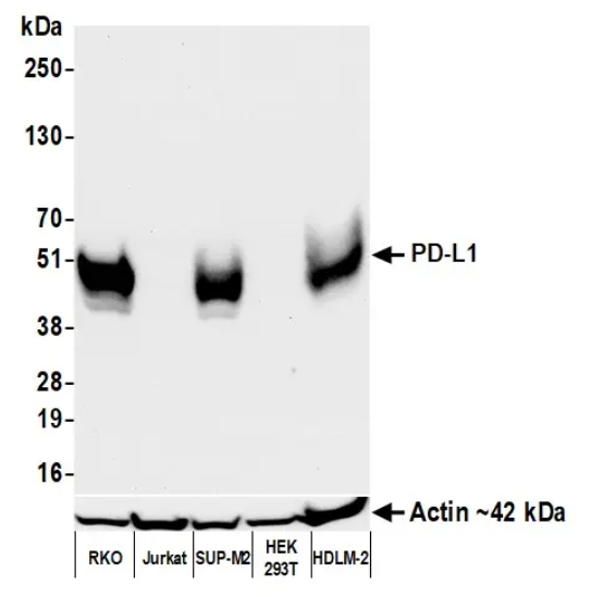
Detection of human PD-L1 by western blot. Samples: Whole cell lysate (50 µg) from RKO, Jurkat, SUP-M2, HEK293T, and HDLM-2 cells prepared using NETN lysis buffer. Antibody: Rabbit anti-PD-L1 recombinant monoclonal antibody used at 1:1000. Secondary: HRP-conjugated goat anti-rabbit IgG. Detection: Chemiluminescence with an exposure time of 30 seconds. Lower Panel: Rabbit anti-Actin recombinant monoclonal antibody
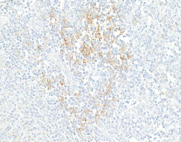
Detection of human PD-L1 by immunohistochemistry. Sample: FFPE section of human tonsil. Antibody: Rabbit anti-PD-L1 recombinant monoclonal antibody. Secondary: HRP-conjugated goat anti-rabbit IgG
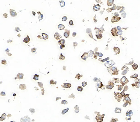
Detection of human PD-L1 by immunocytochemistry. Sample: FFPE section of human SUP-M2 cells. Antibody: Rabbit anti-PD-L1 recombinant monoclonal antibody. Secondary: HRP-conjugated goat anti-rabbit IgG

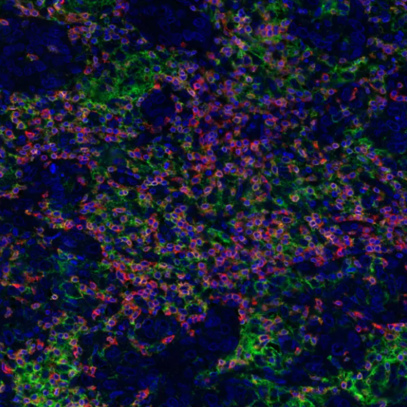
Detection of human PD-L1 (red) and Lamin-A/C (green) in FFPE lung carcinoma by IHC-IF. Antibody: Rabbit anti-PD-L1 recombinant monoclonal and rabbit anti-Lamin-A/C. Secondary: DyLight® 594-conjugated goat anti-rabbit IgG and DyLight® 488-conjugated goat anti-rabbit IgG. Counterstain: DAPI (blue).
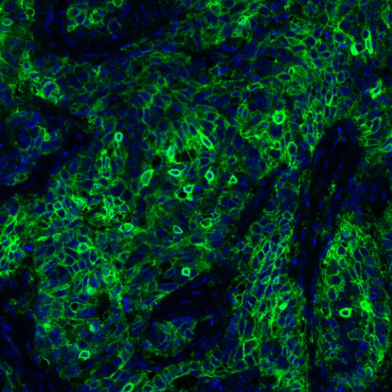
Detection of human PD-L1 (green) by immunohistochemistry. Sample: FFPE section of human lung carcinoma. Antibody: Rabbit anti-PD-L1 recombinant monoclonal antibody used at 1:250. Secondary: HRP-conjugated goat anti-rabbit IgG. Substrate: Opal™. Counterstain: DAPI (blue).
Detection of human PD-L1 by immunocytochemistry. Sample: FFPE section of human SUP-M2 cells. Antibody: Rabbit anti-PD-L1 recombinant monoclonal antibody Secondary: HRP-conjugated goat anti-rabbit IgG
- Item 1 of 5
Recombinant PD-L1 antibody [AC37] [orb1090509]
IHC-P, WB
Human
Rabbit
Monoclonal
Unconjugated
50 μl, 100 μl - Item 1 of 1
Rabbit anti-PD-L1 Recombinant Monoclonal Antibody [orb1519950]
FC, ICC, IF, IHC, IP, WB
Human
Rabbit
Recombinant
Unconjugated
10 μl 
Recombinant PD-L1 / PDCD1LG1 / CD274 / B7-H1 (Cancer Immunotherapy Target) Antibody [orb751369]
IHC
Human
Rabbit
Monoclonal
Unconjugated
100 μgRecombinant PD-L1 / PDCD1LG1 / CD274 / B7-H1 (Cancer Immunotherapy Target) Antibody [orb751310]
FC, IF, IHC, WB
Human, Mouse
Rabbit
Monoclonal
Unconjugated
100 μgRabbit anti-PD-L1 Recombinant Monoclonal Antibody [orb1519951]
FC, ICC, IF, IHC, IP, WB
Human
Rabbit
Recombinant
Unconjugated
100 μg
![Recombinant PD-L1 antibody [AC37]](/images//pub/media/catalog/product/NewWebsite/1/orb1090509_1.png)
![Recombinant PD-L1 antibody [AC37]](/images/pub/media/catalog/product/NewWebsite/1/orb1090509_2.png)
![Recombinant PD-L1 antibody [AC37]](/images/pub/media/catalog/product/NewWebsite/1/orb1090509_3.png)
![Recombinant PD-L1 antibody [AC37]](/images/pub/media/catalog/product/NewWebsite/1/orb1090509_4.png)
![Recombinant PD-L1 antibody [AC37]](/images/pub/media/catalog/product/NewWebsite/1/orb1090509_5.png)

