You have no items in your shopping cart.
Cart summary
Item 1 of 5
Item 1 of 5
Granzyme B Antibody
Catalog Number: orb1519946

| Catalog Number | orb1519946 |
|---|---|
| Category | Antibodies |
| Description | Granzyme B Antibody |
| Species/Host | Rabbit |
| Clonality | Recombinant |
| Clone Number | BLR022E |
| Tested applications | ICC, IF, IHC, WB |
| Reactivity | Human |
| Immunogen | Residues 19-247 (mature protein) |
| Antibody Type | Primary Antibody |
| Concentration | 50 µg/ml |
| Dilution range | Flow-Cyt Fixed in 4% formaldehyde and permeabilized with 90% methanol. 1 µl per 1 x 10^6 cells. Immunocytochemistry (ICC) 1:100 - 1:500. Epitope retrieval with citrate buffer pH6.0 is recommended for FFPE cell sections. Immunohistochemistry (IHC) 1:100 - 1:500. Epitope retrieval with citrate buffer pH 6.0 is recommended for FFPE tissue sections. IHC-IF 1:100 to 1:500. Epitope retrieval with citrate buffer pH6.0 is recommended for FFPE tissue sections. Multiplex Immunofluorescence (mIF) 1:250. Western Blot (WB) 1:1000 |
| Form/Appearance | Whole IgG |
| Purity | The monoclonal antibody was purified from cell culture supernatant |
| Conjugation | Unconjugated |
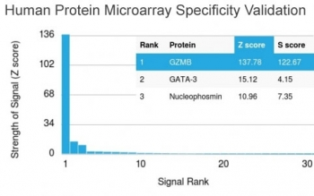
| Target | Granzyme B |
| UniProt ID | P10144 |
| NCBI | NP_004122.2 |
| Storage | 2 - 8°C |
| Buffer/Preservatives | Borate Buffered Saline (BBS) pH 8.2 with 0.1% rAlbumin and 0.09% Sodium Azide |
| Alternative names | C11; cathepsin G-like 1; CCPI; CGL1; CGL-1; CSPB; Read more... |
| Background | Granzyme B is a member of the granzyme subfamily of proteins, part of the peptidase S1 family of serine proteases. The preproprotein is secreted by natural killer (NK) cells and cytotoxic T lymphocytes (CTLs) and proteolytically processed to generate the active protease, which induces target cell apoptosis. This protein also processes cytokines and degrades extracellular matrix proteins, and these roles are implicated in chronic inflammation and wound healing |
| Note | For research use only |
| Application notes | All western blot analysis is performed using 5% Milk-TBST for blocking and as antibody diluent. Primary antibody is incubated overnight |
| Expiration Date | 12 months from date of receipt. |
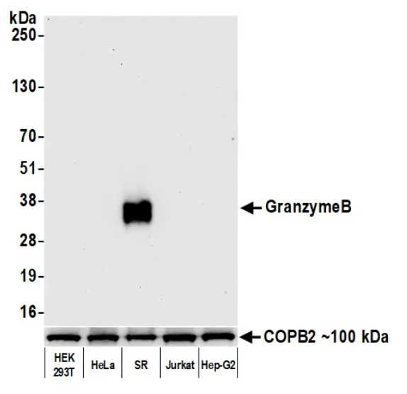
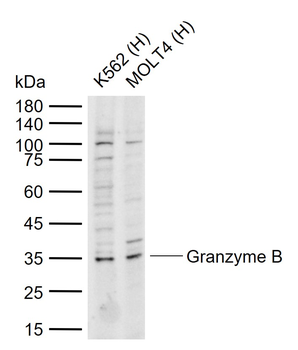
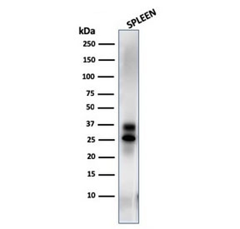
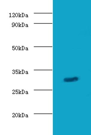
Detection of human Granzyme B by western blot. Samples: Whole cell lysate (15 µg) from HEK293T, HeLa, SR, Jurkat, and Hep-G2 cells prepared using NETN lysis buffer. Antibody: Rabbit anti-GranzymeB recombinant monoclonal antibody used at 1:1000. Secondary: HRP-conjugated goat anti-rabbit IgG. Detection: Chemiluminescence with an exposure time of 10 seconds. Lower Panel: Rabbit anti-COPB2 antibody
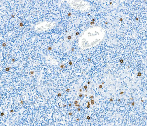
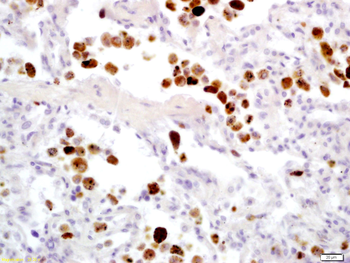
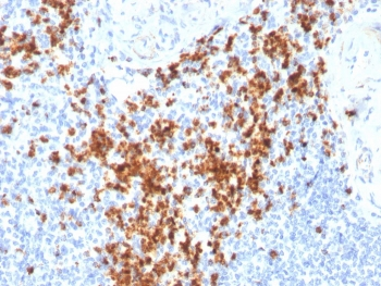
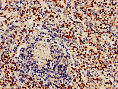
Detection of human Granzyme B by immunohistochemistry. Sample: FFPE section of lymph node. Antibody: Rabbit anti-Granzyme B recombinant monoclonal antibody. Secondary: HRP-conjugated goat anti-rabbit IgG

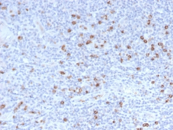
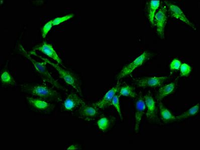
Detection of human Granzyme B by immunocytochemistry. Sample: FFPE section of Granzyme B over-expressing cells (left) and Granzyme H over-expressing cells (right). Antibody: Rabbit anti-Granzyme B recombinant monoclonal antibody. Secondary: HRP-conjugated goat anti-rabbit IgG
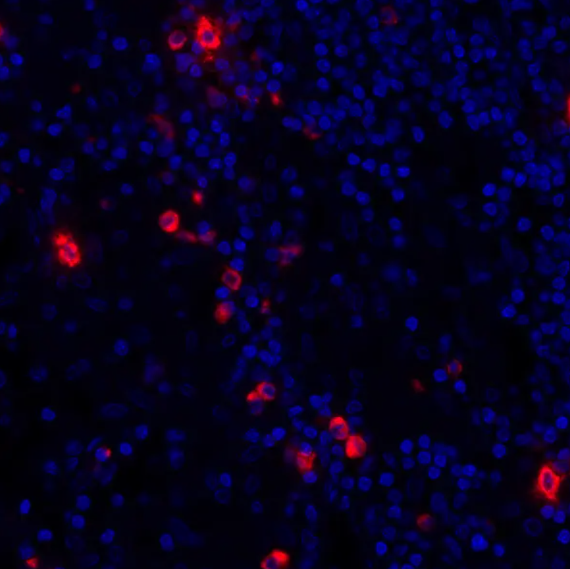
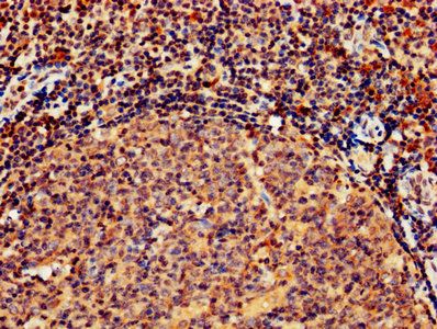
Detection of human Granzyme B (red) by immunohistochemistry. Sample: FFPE section of human lung carcinoma. Antibody: Rabbit anti-Granzyme B recombinant monoclonal antibody used at 1:250. Secondary: HRP-conjugated goat anti-rabbit IgG Substrate: Opal™. Counterstain: DAPI (blue)

Detection of human Granzyme B (shaded) in SR cells by flow cytometry. Antibody: Rabbit anti-Granzyme B recombinant monoclonal antibody or isotype control (unshaded). Secondary: DyLight® 650-conjugated goat anti-rabbit IgG
- Item 1 of 3
Granzyme B Rabbit Polyclonal Antibody [orb10738]
FC, IF, IHC-Fr, IHC-P, WB
Bovine, Mouse, Porcine, Rat
Human
Rabbit
Polyclonal
Unconjugated
50 μl, 100 μl, 200 μl - Item 1 of 5
- Item 1 of 5
- Item 1 of 4
- Item 1 of 3