You have no items in your shopping cart.
Cart summary
Item 1 of 7
Item 1 of 7
Rabbit anti-EGFR Recombinant Monoclonal Antibody
Catalog Number: orb1784586

| Catalog Number | orb1784586 |
|---|---|
| Category | Antibodies |
| Description | Rabbit anti-EGFR Recombinant Monoclonal Antibody |
| Species/Host | Rabbit |
| Clonality | Recombinant |
| Clone Number | BLR252L |
| Tested applications | FC, ICC, IHC, IP, WB |
| Reactivity | Human, Mouse |
| Immunogen | Between 1160 and C-terminus |
| Concentration | 1000 µg/ml |
| Dilution range | WB: 1:1000, IP: 6 µl/mg lysate, IHC: 1:100 to 1:500. Epitope retrieval with citrate buffer pH 6.0 is recommended for FFPE tissue sections, ICC: 1:100 to 1:500. Epitope retrieval with citrate buffer pH 6.0 is recommended for FFPE cell sections |
| Form/Appearance | Whole IgG |
| Conjugation | Unconjugated |
| Target | EGFR |
| UniProt ID | P00533 |
| Storage | Maintain refrigerated at 2-8°C for up to 2 weeks. For long term storage store at -20°C in small aliquots to prevent freeze-thaw cycles. |
| Buffer/Preservatives | Borate Buffered Saline (BBS) pH 8.2 with 0.09% Sodium Azide, rAlbumin-Free |
| Alternative names | erb-b2 receptor tyrosine kinase 1; proto-oncogene Read more... |
| Note | For research use only |
| Application notes | Format: Whole IgG |
| Expiration Date | 12 months from date of receipt. |

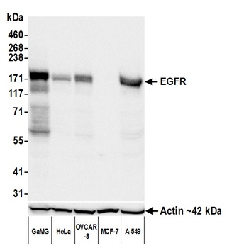
Detection of human EGFR by western blot. Samples: Whole cell lysate (10 µg) from GaMG, HeLa, OVCAR-8, MCF-7, and A-549 cells prepared using NETN lysis buffer. Antibody: Rabbit anti-EGFR recombinant monoclonal antibody (orb1784586) used at 1:1000. Secondary: HRP-conjugated goat anti-rabbit IgG. Detection: Chemiluminescence with an exposure time of 30 seconds. Lower Panel: Rabbit anti-Actin recombinant monoclonal antibody.

Detection of mouse EGFR by western blot. Samples: Whole cell lysate (10 µg) from AML12, NIH 3T3, and mIMCD-3 cells prepared using NETN lysis buffer. Antibody: Rabbit anti-EGFR recombinant monoclonal antibody (orb1784586) used at 1:1000. Secondary: HRP-conjugated goat anti-rabbit IgG. Detection: Chemiluminescence with an exposure time of 30 seconds. Lower Panel: Rabbit anti-Actin recombinant monoclonal antibody.

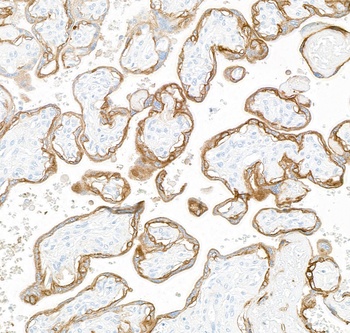
Detection of human EGFR by immunohistochemistry. Sample: FFPE section of human placenta. Antibody: Rabbit anti-EGFR recombinant monoclonal antibody (orb1784586). Secondary: HRP-conjugated goat anti-rabbit IgG.

Detection of mouse EGFR (shaded) in mIMCD-3 cells by flow cytometry. Antibody: Rabbit anti-EGFR recombinant monoclonal antibody (orb1784586) or isotype control (unshaded). Secondary: DyLight® 650-conjugated goat anti-rabbit IgG.

Detection of human EGFR (shaded) in FaDu cells by flow cytometry. Antibody: Rabbit anti-EGFR recombinant monoclonal antibody (orb1784586) or isotype control (unshaded). Secondary: DyLight® 650-conjugated goat anti-rabbit IgG.

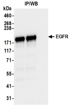
Detection of human EGFR by western blot of immunoprecipitates. Samples: Whole cell lysate (1 mg per IP; 10% of IP loaded) from OVCAR-8 cells prepared using NETN lysis buffer. Antibodies: Rabbit anti-EGFR recombinant monoclonal antibody (orb1784586) used for IP at 6 µl per reaction.

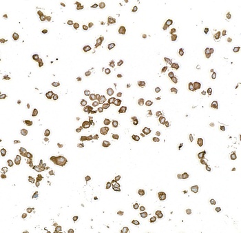
Detection of human EGFR by immunocytochemistry. Sample: FFPE section of A-431 cells. Antibody: Rabbit anti-EGFR recombinant monoclonal antibody (orb1784586). Secondary: HRP-conjugated goat anti-rabbit IgG.
- Item 1 of 7
Rabbit anti-EGFR Recombinant Monoclonal Antibody [orb1784585]
FC, ICC, IHC, IP, WB
Human, Mouse
Rabbit
Recombinant
Unconjugated
10 μl - Item 1 of 4
Rabbit anti-EGFR Recombinant Monoclonal Antibody [orb1710835]
ICC, IHC, IP, WB
Human
Rabbit
Recombinant
Unconjugated
10 μl - Item 1 of 4
Rabbit anti-EGFR Recombinant Monoclonal Antibody [orb1710837]
ICC, IHC, IP, WB
Human
Rabbit
Recombinant
Unconjugated
100 μl 
Rabbit anti-EGFR Recombinant Monoclonal Antibody [orb1710836]
ICC, IHC, IP, WB
Human
Rabbit
Recombinant
Unconjugated
100 μg