You have no items in your shopping cart.
Cart summary
Item 1 of 4
Item 1 of 4
Rabbit anti-CD8 alpha Recombinant Monoclonal Antibody
Catalog Number: orb1519887

| Catalog Number | orb1519887 |
|---|---|
| Category | Antibodies |
| Description | CD8 alpha Antibody |
| Species/Host | Rabbit |
| Clonality | Recombinant |
| Clone Number | BLR044F |
| Tested applications | FC, IF, IHC, WB |
| Reactivity | Human |
| Immunogen | Between 185 and C-terminus |
| Antibody Type | Primary Antibody |
| Concentration | 250 µg/ml |
| Dilution range | Flow-Cyt Fixed in 4% formaldehyde and permeabilized with 90% methanol. 0.5 µl per 1 x 10^6 cells. Immunohistochemistry (IHC) 1:100 to 1:500. Epitope retrieval with citrate buffer pH6.0 is recommended for FFPE tissue sections. IHC-IF 1:100 to 1:500. Epitope retrieval with citrate buffer pH6.0 is recommended for FFPE cell sections. Multiplex Immunofluorescence (mIF) 1:250. Western Blot (WB) 1:1000 |
| Form/Appearance | Whole IgG |
| Purity | Recombinant antibody was purified from cell culture supernatant |
| Conjugation | Unconjugated |
| Target | CD8 alpha |
| UniProt ID | P01732 |
| NCBI | NP_001759.3 |
| Storage | 2 - 8°C |
| Buffer/Preservatives | Borate Buffered Saline (BBS) pH 8.2 with 0.1% rAlbumin and 0.09% Sodium Azide |
| Alternative names | Leu2 T-lymphocyte antigen; T-lymphocyte differenti Read more... |
| Background | The CD8 antigen is a cell surface glycoprotein found on most cytotoxic T lymphocytes that mediates efficient cell-cell interactions within the immune system. The CD8 antigen acts as a coreceptor with the T-cell receptor on the T lymphocyte to recognize antigens displayed by an antigen presenting cell in the context of class I MHC molecules. The coreceptor functions as either a homodimer composed of two alpha chains or as a heterodimer composed of one alpha and one beta chain. Both alpha and beta chains share significant homology to immunoglobulin variable light chains. This gene encodes the CD8 alpha chain |
| Note | For research use only |
| Application notes | Format: Whole IgG |
| Expiration Date | 12 months from date of receipt. |
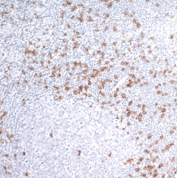
Detection of human CD8 alpha by immunohistochemistry. Sample: FFPE section of human tonsil. Antibody: Rabbit anti-CD8 alpha recombinant monoclonal antibody used at 1:250. Secondary: HRP-conjugated goat anti-rabbit IgG). Substrate: DAB
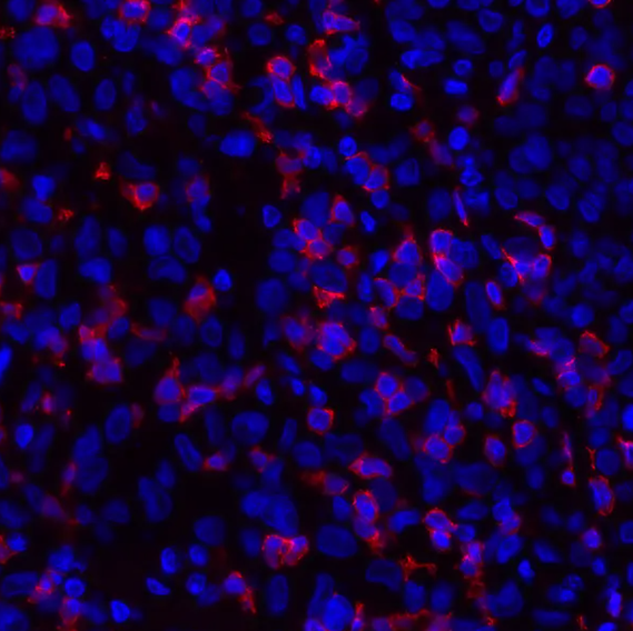
Detection of human CD8 alpha by immunhistochemistry. Sample: FFPE section of human melanoma. Antibody: Rabbit anti-CD8 alpha recombinant monoclonal antibody used at 1:100. Secondary: DyLight® 594-conjugated goat anti-rabbit IgG
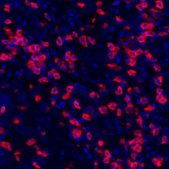
Detection of human CD8 alpha (red) by immunohistochemistry. Sample: FFPE section of human breast carcinoma. Antibody: Rabbit anti-CD8 alpha recombinant monoclonal antibody used at 1:250. Secondary: HRP-conjugated goat anti-rabbit IgG. Substrate: Opal™. Counterstain: DAPI (blue).
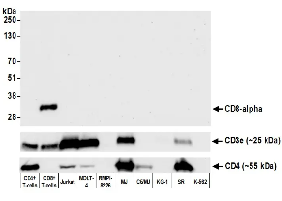
Detection of human CD8 alpha by western blot. Samples: Whole cell lysate (50 µg) from CD4+ T-cells, CD8+ T-cells, Jurkat, MOLT-4, RPMI-8226, MJ, C5/MJ, KG-1, SR, and K-562 cells prepared using NETN lysis buffer. Antibody: Rabbit anti-CD8 alpha recombinant monoclonal antibody used at 1:1000. Secondary: HRP-conjugated goat anti-rabbit IgG. Detection: Chemiluminescence with an exposure time of 30 seconds. Lower panel: Recombinant monoclonal antibodies to CD3e and CD4
- Item 1 of 1
Rabbit anti-CD8 alpha Recombinant Monoclonal Antibody [orb1519885]
FC, IF, IHC, WB
Human
Rabbit
Recombinant
Unconjugated
10 μl 
Recombinant CD8A (Cytotoxic / Suppressor T-Cell Marker) Antibody [orb751347]
ELISA, FC
Human, Primate
Rabbit
Monoclonal
Unconjugated
100 μgRecombinant CD8A (Cytotoxic / Suppressor T-Cell Marker) Antibody [orb751419]
IHC
Human
Rabbit
Monoclonal
Unconjugated
100 μgRabbit anti-CD8 alpha Recombinant Monoclonal Antibody [orb1519886]
IF, IHC, WB
Human
Rabbit
Recombinant
Unconjugated
100 μgRabbit anti-CD8 alpha Recombinant Monoclonal Antibody [orb1519610]
FC, IHC, WB
Human
Rabbit
Recombinant
Unconjugated
10 μl

