You have no items in your shopping cart.
Cart summary
Item 1 of 3
Item 1 of 3
PTGS1 Antibody (C-term)
Catalog Number: orb1931423

| Catalog Number | orb1931423 |
|---|---|
| Category | Antibodies |
| Description | Purified Rabbit Polyclonal Antibody (Pab) |
| Species/Host | Rabbit |
| Clonality | Polyclonal |
| Clone Number | RB17407 |
| Tested applications | IF, IHC-P, WB |
| Reactivity | Human |
| Isotype | Rabbit IgG |
| Dilution range | IF: 1:10~50, WB: 1:1000, IHC-P: 1:10~50 |
| Form/Appearance | Purified polyclonal antibody supplied in PBS with 0.09% (W/V) sodium azide. This antibody is prepared by Saturated Ammonium Sulfate (SAS) precipitation followed by dialysis against PBS. |
| Conjugation | Unconjugated |
| MW | 68686 Da |
| Target | This PTGS1 antibody is generated from rabbits immunized with a KLH conjugated synthetic peptide between 571-599 amino acids from the C-terminal region of human PTGS1. |
| UniProt ID | P23219 |
| NCBI | NP_000953.2, NP_001258095.1, NP_542158.1, NP_001258297.1, NP_001258094.1 |
| Storage | Maintain refrigerated at 2-8°C for up to 2 weeks. For long term storage store at -20°C in small aliquots to prevent freeze-thaw cycles |
| Alternative names | Prostaglandin G/H synthase 1, Cyclooxygenase-1, CO Read more... |
| Note | For research use only |
| Expiration Date | 12 months from date of receipt. |

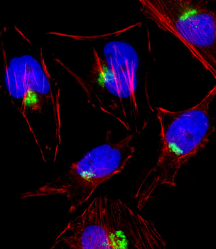
Fluorescent image of Hela cell stained with PTGS1 Antibody (C-term).Hela cells were fixed with 4% PFA (20 min), permeabilized with Triton X-100 (0.1%, 10 min), then incubated with PTGS1 primary antibody (1:25, 1 h at 37°C). For secondary antibody, Alexa Fluor 488 conjugated donkey anti-rabbit antibody (green) was used (1:400, 50 min at 37°C).Cytoplasmic actin was counterstained with Alexa Fluor 555 (red) conjugated Phalloidin (7units/ml, 1 h at 37°C).PTGS1 immunoreactivity is localized to Golgi significantly.
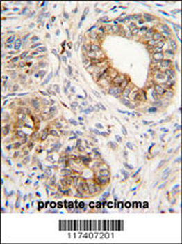
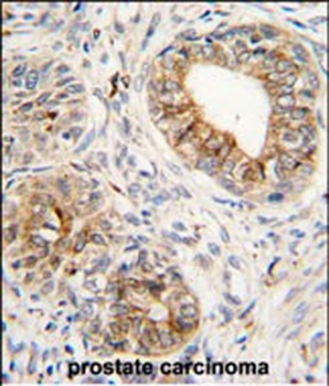
Formalin-fixed and paraffin-embedded human prostata carcinoma tissue reacted with PTGS1 antibody (C-term), which was peroxidase-conjugated to the secondary antibody, followed by DAB staining. This data demonstrates the use of this antibody for immunohistochemistry; clinical relevance has not been evaluated.
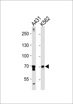
PTGS1 Antibody (C-term) western blot analysis in A431, K562 cell line lysates (35 ug/lane). This demonstrates the PTGS1 antibody detected the PTGS1 protein (arrow).
- Item 1 of 3