You have no items in your shopping cart.
Cart summary
Item 1 of 4
Item 1 of 4
PSD95 Antibody: APC
Catalog Number: orb147023

| Catalog Number | orb147023 |
|---|---|
| Category | Antibodies |
| Description | Mouse monoclonal to PSD95 (APC). Postsynaptic Density protein 95 (PSD95), also known as Postsynaptic Density protein 95 (PSD95), also known as... |
| Species/Host | Mouse |
| Clonality | Monoclonal |
| Clone Number | 7000 |
| Tested applications | ICC, IF, IHC |
| Reactivity | Bovine, Human, Mouse, Rat |
| Isotype | IgG1 |
| Immunogen | Recombinant rat PSD-95 |
| Concentration | 1 mg/ml |
| Dilution range | WB (1:1000), IHC (1:1000), ICC/IF (1:100) |
| Conjugation | APC |
| MW | 75kDa |
| Target | PSD95 |
| Entrez | 29495 |
| UniProt ID | P31016 |
| NCBI | NP_062567.1 |
| Storage | Conjugated antibodies should be stored according to the product label |
| Buffer/Preservatives | 95.64mM Phosphate, 2.48mM MES and 2mM EDTA |
| Alternative names | PSD 95 antibody, PSD-95 antibody, DLG4 antibody, S Read more... |
| Note | For research use only |
| Application notes | 1 µg/ml was sufficient for detection of PSD-95 on 20 µg rat brain tissue extract by ECL immunoblot analysis using Goat Anti-Mouse IgG: HRP as the secondary. |
| Expiration Date | 12 months from date of receipt. |

Immunohistochemistry analysis using Mouse Anti-PSD95 Monoclonal Antibody, Clone 7E3. Tissue: Neocortex. Species: Rat. Primary Antibody: Mouse Anti-PSD95 Monoclonal Antibody at 1:1000.

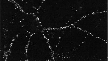
Immunocytochemistry/Immunofluorescence analysis using Mouse Anti-PSD95 Monoclonal Antibody, Clone 7E3. Tissue: HaCaT cells. Species: Human. Fixation: Cold 100% methanol for 10 minutes at -20°C. Primary Antibody: Mouse Anti-PSD95 Monoclonal Antibody at 1:100 for 1 hour at RT. Secondary Antibody: FITC Goat Anti-Mouse (green) at 1:50 for 1 hour at RT. Localization: Filamentous-like staining.
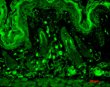
Immunohistochemistry analysis using Mouse Anti-PSD95 Monoclonal Antibody, Clone 7E3. Tissue: backskin. Species: Mouse. Fixation: Bouin's Fixative and paraffin-embedded. Primary Antibody: Mouse Anti-PSD95 Monoclonal Antibody at 1:100 for 1 hour at RT. Secondary Antibody: FITC Goat Anti-Mouse (green) at 1:50 for 1 hour at RT. Localization: Basal cell staining in the epidermis, some hair follicle staining, dermal staining. Backskin obtained from transgenic mice.
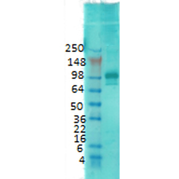
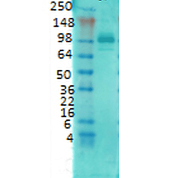
Western Blot analysis of Rat brain membrane lysate showing detection of PSD95 protein using Mouse Anti-PSD95 Monoclonal Antibody, Clone 7E3. Primary Antibody: Mouse Anti-PSD95 Monoclonal Antibody at 1:1000.
- Item 1 of 3

PSD95 Rabbit Polyclonal Antibody (APC) [orb999673]
FC, IF
Equine, Porcine, Rabbit, Sheep
Human, Mouse, Rat
Rabbit
Polyclonal
APC
100 μlPSD95 Rabbit Polyclonal Antibody (APC) [orb999674]
IF
Bovine, Canine, Equine, Human, Porcine, Rabbit, Sheep
Mouse, Rat
Rabbit
Polyclonal
APC
100 μlGDA/GDC Rabbit Polyclonal Antibody (APC) [orb997442]
ICC, IF
Bovine, Canine, Equine, Human, Mouse, Rabbit, Rat
Rabbit
Polyclonal
APC
100 μlDLGAP1 Rabbit Polyclonal Antibody (APC) [orb1005167]
ICC, IF
Bovine, Canine, Equine, Human, Mouse, Porcine, Rabbit, Rat, Sheep
Rabbit
Polyclonal
APC
100 μl