You have no items in your shopping cart.
Cart summary
Item 1 of 3
Item 1 of 3
PMEL17 / Melanoma gp100 Antibody
Catalog Number: orb749784

| Catalog Number | orb749784 |
|---|---|
| Category | Antibodies |
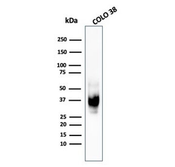
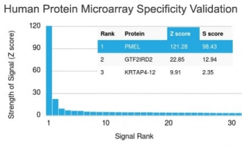
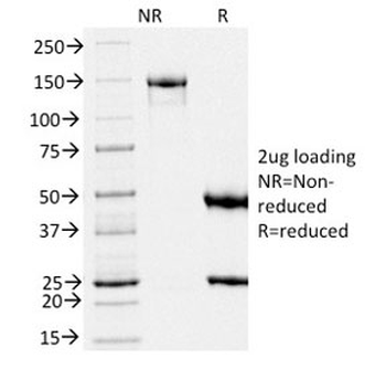
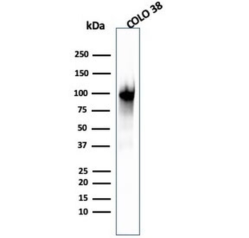
| Description | Cytotoxic T lymphocytes (CTL's) recognize melanoma-associated antigens, which belong to three main groups. These groups include tumor-associated testis-specific antigens, melanocyte differentiation antigens and mutated or aberrantly expressed antigens, which are routinely used as markers to identify melanomas based on their binding to specific monoclonal antibodies. gp100, also designated ME20-M, ME20-S and PMEL 17, is classified as a melanocyte differentiation antigen and is expressed at low levels in normal cell lines and tissues, but is upregulated in melanocytes. gp100 is a highly glycosylated protein. It is also the product of proteolytic cleavage, which results in a secreted protein.The gp100 molecule is a 100kDa glycosylated protein that is cleaved into a small (26kDa) carboxy-terminal fragment and a larger amino- terminal section (60-64 kDa), which is subsequently cleaved to generate 26kDa and 34-38kDa fragments. |
| Species/Host | Mouse |
| Clonality | Monoclonal |
| Clone Number | PMEL/783 |
| Tested applications | IHC-P, WB |
| Reactivity | Human |
| Isotype | Mouse IgG1, kappa |
| Immunogen | Recombinant human protein was used as the immunogen for the PMEL17 antibody. |
| Dilution range | Western blot: 1-2ug/ml,Immunohistochemistry (FFPE): 1-2ug/ml for 30 min at RT (1) (2) |
| Purity | Protein G affinity chromatography |
| Conjugation | Unconjugated |
| Formula | 0.2 mg/ml in 1X PBS with 0.1 mg/ml BSA (US sourced) and 0.05% sodium azide |
| Hazard Information | This PMEL17 antibody is available for research use only. |
| UniProt ID | P40967 |
| Storage | Store the PMEL17 antibody at 2-8°C (with azide) or aliquot and store at -20°C or colder (without azide). |
| Buffer/Preservatives | 0.2 mg/ml in 1X PBS with 0.1 mg/ml rAlbumin (US sourced) and 0.05% sodium azide |
| Note | For research use only |
| Application notes | Optimal dilution of the PMEL17 antibody should be determined by the researcher.1. Staining of formalin-fixed tissues requires boiling tissue sections in pH 9 10mM Tris with 1mM EDTA for 10-20 min followed by cooling at RT for 20 min.2. The prediluted format is supplied in a dropper bottle and is optimized for use in IHC. After epitope retrieval step (if required), drip mAb solution onto the tissue section and incubate at RT for 30 min. |
| Expiration Date | 12 months from date of receipt. |
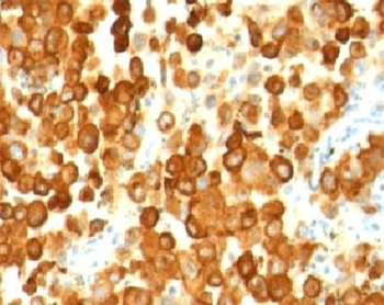
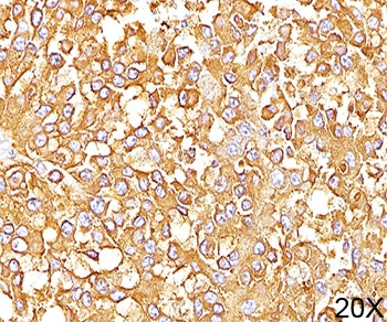
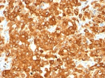
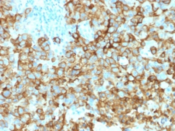
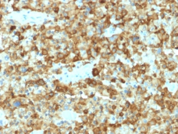
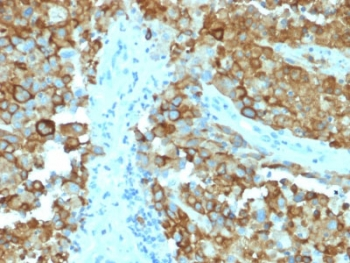
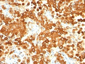
IHC: Formalin-fixed, paraffin-embedded human melanoma stained with PMEL17 antibody (PMEL/783).
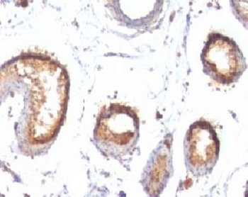
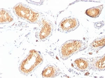
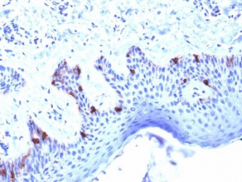
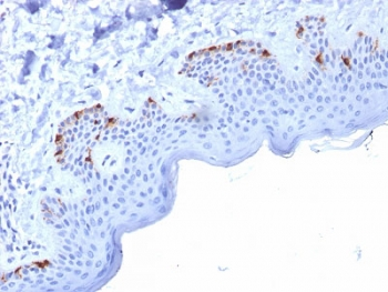
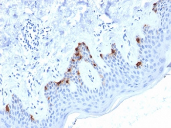
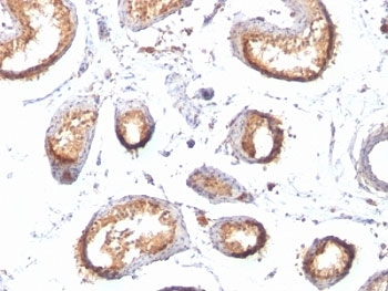
IHC: Formalin-fixed, paraffin-embedded human testis stained with PMEL17 antibody (PMEL/783).
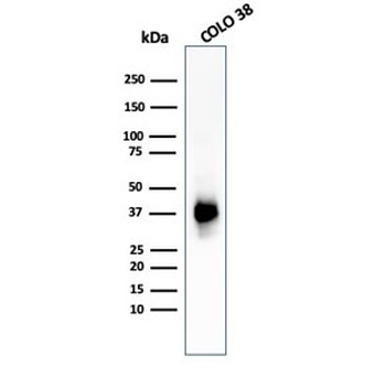
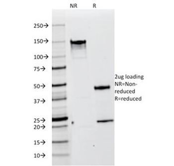
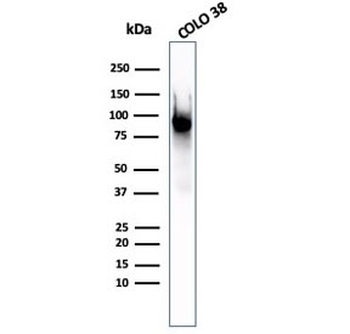
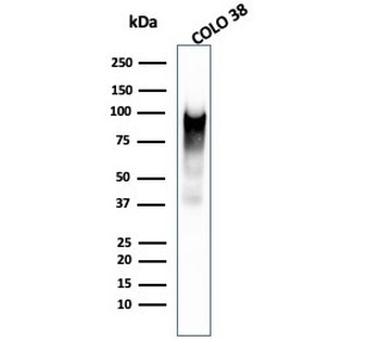
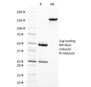
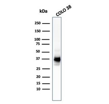
Western blot testing of human COLO-38 cell lysate with PMEL17 antibody (clone PMEL/783).
- Item 1 of 6
PMEL17 / Melanoma gp100 Antibody [orb248538]
FACS, IF, IHC-P, WB
Human
Mouse
Monoclonal
Unconjugated
100 μg, 20 μg - Item 1 of 5
PMEL17 / Melanoma gp100 Antibody [orb606652]
ELISA, IHC-P, WB
Human
Mouse
Monoclonal
Unconjugated
100 μg, 20 μg - Item 1 of 5
PMEL17 / Melanoma gp100 Antibody [orb606653]
ELISA, IHC-P, WB
Human
Mouse
Monoclonal
Unconjugated
100 μg, 20 μg - Item 1 of 5
PMEL17 / Melanoma gp100 Antibody [orb606654]
ELISA, IHC-P, WB
Human
Mouse
Monoclonal
Unconjugated
100 μg, 20 μg - Item 1 of 3
PMEL17 / Melanoma gp100 Antibody Cocktail [orb749785]
IHC-P, WB
Human
Mouse
Monoclonal
Unconjugated
20 μg, 100 μg
Submit a review
Filter by Rating

- 5 stars

- 4 stars
- 3 stars
- 2 stars
- 1 stars
