You have no items in your shopping cart.
Cart summary
Item 1 of 4
Item 1 of 4
PIN1 antibody
Catalog Number: orb345588

| Catalog Number | orb345588 |
|---|---|
| Category | Antibodies |
| Description | PIN1 antibody |
| Species/Host | Rabbit |
| Clonality | Polyclonal |
| Tested applications | ELISA, IHC, IP, WB |
| Reactivity | Human |
| Isotype | IgG |
| Immunogen | This affinity purified antibody was prepared from whole rabbit serum produced by repeated immunizations with a synthetic peptide corresponding to an internal sequence of human Pin1. |
| Concentration | 1.1 mg/mL |
| Dilution range | ELISA: 1:2,500 - 1:10,000, IHC: 5µg/mL, IP: 1:100, WB: 1:500 - 1:3,000 |
| Form/Appearance | Liquid (sterile filtered) |
| Purity | This affinity purified antibody is directed against human Pin1. The product was affinity purified from monospecific antiserum by immunoaffinity chromatography. A BLAST analysis was used to suggest cross-reactivity with Pin1 from human, dog, bovine and monkey based on a 100% homology with the immunizing sequence. Expect partial reactivity with Pin1 from mouse and rat sources based on 92% sequence homologies. Reactivity against homologues from other sources is not known. |
| Conjugation | Unconjugated |
| UniProt ID | Q13526 |
| NCBI | 5453898 |
| Storage | Store vial at -20° C prior to opening. Aliquot contents and freeze at -20° C or below for extended storage. Avoid cycles of freezing and thawing. Centrifuge product if not completely clear after standing at room temperature. This product is stable for several weeks at 4° C as an undiluted liquid. Dilute only prior to immediate use. |
| Buffer/Preservatives | 0.01% (w/v) Sodium Azide |
| Alternative names | rabbit anti-PIN! antibody, PIN-1, PIN 1, Peptidyl- Read more... |
| Note | For research use only |
| Application notes | This affinity purified antibody has been tested for use in ELISA, Immunohistochemistry, and western blotting. Specific conditions for reactivity should be optimized by the end user. Expect a band approximately 18 kDa in size corresponding to Pin1 by western blotting in the appropriate cell lysate or extract. Lysates from 3T3, Jurkat, 293 or HeLa cells, as well as HeLa nuclear extract, are recommended for use as positive controls. |
| Expiration Date | 12 months from date of receipt. |
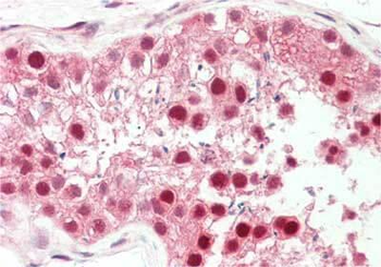
Immunohistochemistry of rabbit anti-PIN1 antibody. Tissue: testis. Fixation: formalin fixed paraffin embedded. Antigen retrieval: not required. Primary antibody: Anti-PIN1 at 5 µg/mL for 1 h at RT. Secondary antibody: Peroxidase rabbit secondary antibody at 1:10000 for 45 min at RT. Staining: PIN-1 as precipitated red signal with hematoxylin purple nuclear counterstain.
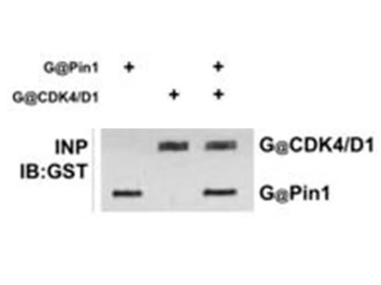
Immunoprecipitation of Rabbit anti-PIN1 antibody. Lane 1: T98G cells incubated with GST-Pin1. Lane 2: T98G cells incubated with GST-CDK4/cyclinD1. Lane 3: T98G cells incubated with GST-Pin1 and GST-CDK4/cyclinD1. Immunoprecipitated with pRb antibody. Load: 25 µg per lane. Primary antibody: anti-GST 1:400 for overnight at 4°C. Secondary antibody: IRDye800™ secondary antibody at 1:10000 for 45 min at RT. Block: 5% BLOTTO overnight at 4°C.
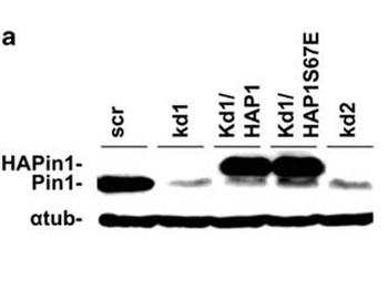
Western Blot of Rabbit anti-PIN1 antibody. Lane 1: T98G cells treated with scrambled (scr). Lane 2: T98G cells treated with PIN1 kd1 shRNA. Lane 3: T98G cells treated with PIN1-overexpressing plasmid HAP1. Lane 4: T98G cells treated with PIN1-overexpressing plasmid HAP1S67E. Lane 5: T98G cells treated with PIN1 kd2 shRNA. Load: 25 µg per lane. Primary antibody: PIN 1 antibody at 1:400 for overnight at 4°C. Secondary antibody: IRDye800™ rabbit secondary antibody at 1:10000 for 45 min at RT. Block: 5% BLOTTO overnight at 4°C. Predicted size: 18 kDa for PIN-1. Other band(s): normalized with a-tubulin (α-tub) antibody.
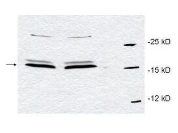
Western blot using Biorbyt's affinity purified anti-Pin1 antibody to detect endogenous Pin1 in HeLa whole cell lysates. The sample was run in duplicate. A band representing Pin1 is indicated by the arrowhead. Cell lysates were electrophoresed using a straight 15% polyacrylamide gel, followed by transfer to nitrocellulose. The membrane was probed with the primary antibody at a 1:700 dilution. A 1:5000 dilution of HRP Gt-a-Rabbit IgG (orb347654) was used with a 15 sec exposure time.
- Item 1 of 5
- Item 1 of 4
PIN1 Antibody [orb1564431]
ICC, IHC-Fr, IHC-P, IP, WB
Hamster, Human, Mouse, Rat
Rabbit
Monoclonal
Unconjugated
100 μl, 50 μl, 20 μl - Item 1 of 4
- Item 1 of 3
PIN1 (phospho-S16) antibody [orb214400]
IF, IH, WB
Human, Mouse, Porcine, Primate, Rat, Zebrafish
Rabbit
Polyclonal
Unconjugated
200 μl, 100 μl, 30 μl - Item 1 of 3


Submit a review
Filter by Rating

- 5 stars

- 4 stars
- 3 stars
- 2 stars
- 1 stars
