You have no items in your shopping cart.
Cart summary
Item 1 of 2
Item 1 of 2
Piccolo Antibody: FITC
Catalog Number: orb147926

| Catalog Number | orb147926 |
|---|---|
| Category | Antibodies |
| Description | Mouse monoclonal to Piccolo (FITC). Piccolo, also referred to as Aczonin, is a large protein which consists of an N-terminal Zn2+ finger, several piccolo-bassoon homology domains and C-terminal PDZ and C2 domains. In general it is found together with bassoon, a related huge multi-domain protein of the CAZ (cytoskeletal matric assembled at active zones). Piccolo is a scaffolding protein for proteins involved in endo- and exocytosis of synaptic vesicles. Piccolo has also been shown to interfere with clathrin medicated endocytosis by binding to the F-actin and dynamin binding protein Abp1.. |
| Species/Host | Mouse |
| Clonality | Monoclonal |
| Clone Number | 6H9-B6 |
| Tested applications | ICC, IF, IHC |
| Reactivity | Human, Mouse, Rat |
| Isotype | IgG2b Kappa |
| Immunogen | Full length recombinant protein (GenBank ID: BC001304.1) |
| Concentration | 1 mg/ml |
| Dilution range | WB (1:1000), ICC/IF (1:100) |
| Conjugation | FITC |
| MW | 550kDa |
| Target | Piccolo |
| Entrez | 26875 |
| UniProt ID | Q9QYX7 |
| NCBI | NP_001104266.1 |
| Storage | Conjugated antibodies should be stored according to the product label |
| Buffer/Preservatives | 640.91mM DMSO, 136.36mM Ethanolamine, 9.09mM Sodium Bicarbonate in 90.9% PBS |
| Alternative names | ACZ antibody, Aczonin antibody, PCLO antibody, Pic Read more... |
| Note | For research use only |
| Application notes | 1 µg/ml of SMC-188 was sufficient for detection of Piccolo in 20 µg of rat brain tissue lysate by colorimetric immunoblot analysis using goat IgG:HRP as the secondary antibody. |
| Expiration Date | 12 months from date of receipt. |

Immunocytochemistry/Immunofluorescence analysis using Mouse Anti-Piccolo Monoclonal Antibody, Clone 6H9-B6. Tissue: Neuroblastoma cell line (SK-N-BE). Species: Human. Fixation: 4% Formaldehyde for 15 min at RT. Primary Antibody: Mouse Anti-Piccolo Monoclonal Antibody at 1:100 for 60 min at RT. Secondary Antibody: Goat Anti-Mouse ATTO 488 at 1:100 for 60 min at RT. Counterstain: Phalloidin Texas Red F-Actin stain; DAPI (blue) nuclear stain at 1:1000, 1:5000 for 60min RT, 5min RT. Localization: Cytoplasm, Endoplasmic Reticulum, Nucleus. Magnification: 60X. (A) DAPI (blue) nuclear stain. (B) Phalloidin Texas Red F-Actin stain. (C) Piccolo Antibody. (D) Composite.
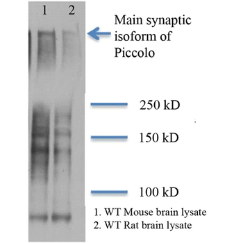
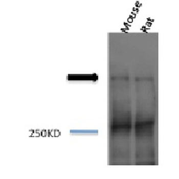
Western Blot analysis of Rat and mouse brain lysates showing detection of Piccolo protein using Mouse Anti-Piccolo Monoclonal Antibody, Clone 6H9-B6. Primary Antibody: Mouse Anti-Piccolo Monoclonal Antibody at 1:1000. (1) Mouse brain lysate; (2) Rat brain lysate.
- Item 1 of 2