You have no items in your shopping cart.
Cart summary
Item 1 of 8
Item 1 of 8
Phospho-p38 MAPK (Thr180 + Tyr182) Rabbit Polyclonal Antibody
Catalog Number: orb6578

| Catalog Number | orb6578 |
|---|---|
| Category | Antibodies |
| Description | Phospho-p38 MAPK (Thr180 + Tyr182) Rabbit Polyclonal Antibody |
| Species/Host | Rabbit |
| Clonality | Polyclonal |
| Tested applications | ELISA, FC, ICC, IF, IHC-Fr, IHC-P, WB |
| Predicted Reactivity | Canine, Equine, Gallus, Porcine, Rat |
| Reactivity | Human, Mouse, Rabbit |
| Isotype | IgG |
| Immunogen | KLH conjugated Synthesised phosphopeptide derived from human p38 MAPK around the phosphorylation site of Thr180/Tyr182 EM(p-T)G(p-Y)VA |
| Concentration | 1mg/ml |
| Dilution range | WB=1:500-2000, IHC-P=1:100-500, IHC-F=1:100-500, ICC/IF=1:100, IF=1:100-500, Flow-Cyt=1μg /test, ELISA=1:5000-10000 |
| Form/Appearance | Liquid |
| Conjugation | Unconjugated |
| MW | 41 kDa |
| Target | MAPK14 |
| UniProt ID | Q16539 |
| RRID | AB_10931075 |
| Storage | Maintain refrigerated at 2-8°C for up to 2 weeks. For long term storage store at -20°C in small aliquots to prevent freeze-thaw cycles. |
| Buffer/Preservatives | 0.01M TBS (pH7.4) with 1% rAlbumin, 0.02% Proclin300 and 50% Glycerol. |
| Alternative names | p38 (phospho T180 + Y182); p-p38 (phospho T180 + Y Read more... |
| Note | For research use only |
| Expiration Date | 12 months from date of receipt. |
Sarin, Navin et al. Key Players of Cisplatin Resistance: Towards a Systems Pharmacology Approach Int J Mol Sci, 19, E767 (2018)
Fan, Lei et al. Astragalus polysaccharide ameliorates lipopolysaccharide-induced cell injury in ATDC5 cells via miR-92a/KLF4 mediation Biomed. Pharmacother., 118, 109180 (2019)

Blank control (blue): HepG2 (fixed with 2% paraformaldehyde for 10 min at 37°C). Primary Antibody: Rabbit Anti-phospho-p38 MAPK (Thr180 + Tyr182) antibody (orb6578, Green), dilution: 1 µg in 100 µl 1X PBS containing 0.5% BSA, Isotype Control Antibody: Rabbit IgG (orange), used under the same conditions, Secondary Antibody: Goat anti-rabbit IgG-FITC (white blue), dilution: 1:200 in 1X PBS containing 0.5% BSA.
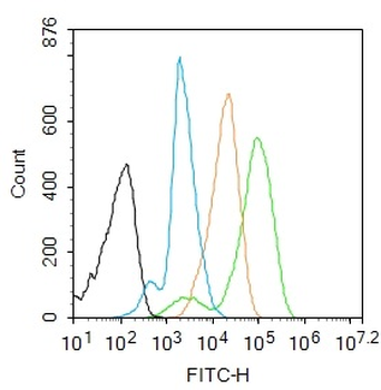
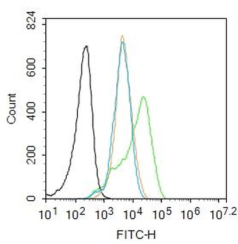
Blank control: Raw264.7. Primary Antibody (green line): Rabbit Anti-phospho-p38 MAPK (Thr180 + Tyr182) antibody (orb6578), dilution: 1 µg/10^6 cells, Isotype Control Antibody (orange line): Rabbit IgG. Secondary Antibody: Goat anti-rabbit IgG-AF488, dilution: 1 µg/Test. Protocol, The cells were fixed with 4% PFA (10 min at room temperature) and then permeabilized with 90% ice-cold methanol for 20 min at -20°C. The cells were then incubated in 5% BSA to block non-specific protein-protein interactions for 30 min at room temperature. Cells stained with Primary Antibody for 30 min at room temperature. The secondary antibody used for 40 min at room temperature. Acquisition of 20000 events was performed.
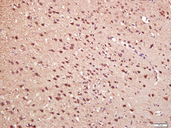
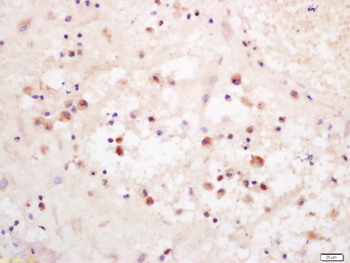
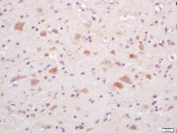
Paraformaldehyde-fixed, paraffin embedded (mouse brain), Antigen retrieval by boiling in sodium citrate buffer (pH6.0) for 15 min, Block endogenous peroxidase by 3% hydrogen peroxide for 20 minutes, Blocking buffer (normal goat serum) at 37°C for 30 min, Antibody incubation with (phospho-p38 MAPK (Thr180 + Tyr182)) Polyclonal Antibody, Unconjugated (orb6578) at 1:200 overnight at 4°C, followed by operating according to SP Kit (Rabbit) instructionsand DAB staining.
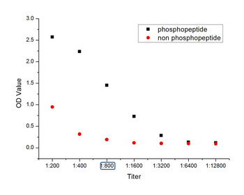
phosphopeptide, non phosphopeptide.
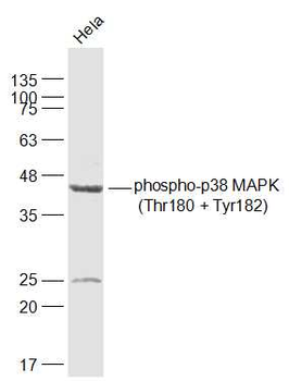
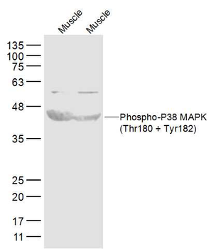
Sample: Hela (Human) Cell Lysate at 30 ug, Primary: Anti-phospho-p38 MAPK (Thr180 + Tyr182) (orb6578) at 1/1000 dilution, Secondary: IRDye800CW Goat Anti-Rabbit IgG at 1/20000 dilution, Predicted band size: 41 kD, Observed band size: 41 kD.
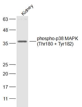
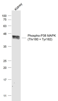
Sample: Kidney (Mouse) Lysate at 40 ug, Primary: Anti-phospho-p38 MAPK (Thr180 + Tyr182) (orb6578) at 1/300 dilution, Secondary: IRDye800CW Goat Anti-Rabbit IgG at 1/20000 dilution, Predicted band size: 41 kD, Observed band size: 41 kD.
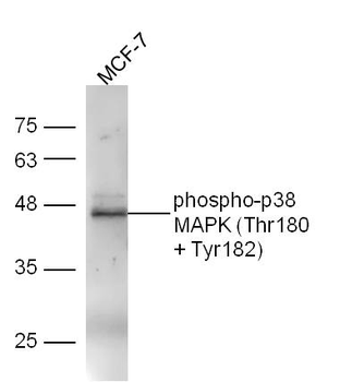
Sample: Mcf-7 Cell Lysate at 30 ug, Primary: Anti-phospho-p38 MAPK (Thr180 + Tyr182) (orb6578) at 1/300 dilution, Secondary: IRDye800CW Goat Anti-Rabbit IgG at 1/20000 dilution, Predicted band size: 41 kD, Observed band size: 45 kD.
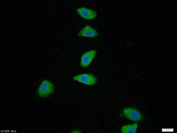
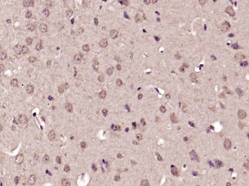
Tissue/Cell: Hela cell, 4% Paraformaldehyde-fixed, Triton X-100 at room temperature for 20 min, Blocking buffer (normal goat serum) at 37°C for 20 min, Antibody incubation with (phospho-p38 MAPK (Thr180 + Tyr182)) polyclonal Antibody, Unconjugated (orb6578) 1:100, 90 minutes at 37°C, followed by a FITC conjugated Goat Anti-Rabbit IgG antibody at 37°C for 90 minutes, DAPI (blue) was used to stain the cell nuclei.
- Item 1 of 8
Phospho-P38 MAPK (Thr180 + Tyr182) Rabbit Polyclonal Antibody [orb11208]
FC, IF, IHC-Fr, IHC-P, WB
Canine, Rabbit
Human, Mouse, Rat
Rabbit
Polyclonal
Unconjugated
50 μl, 100 μl, 200 μl 
Phospho-p38 MAPK (Thr180 + Tyr182) Rabbit Polyclonal Antibody (PE-Cy5) [orb889214]
FC, ICC, IF
Canine, Equine, Gallus, Porcine, Rat
Human, Mouse, Rabbit
Rabbit
Polyclonal
PE/Cy5
100 μlPhospho-p38 MAPK (Thr180 + Tyr182) Rabbit Polyclonal Antibody (BF750) [orb1593745]
FC, ICC, IF
Canine, Equine, Gallus, Porcine, Rat
Human, Mouse, Rabbit
Rabbit
Polyclonal
BF750
100 μlPhospho-p38 MAPK (Thr180 + Tyr182) Rabbit Polyclonal Antibody (BF680) [orb1593746]
FC, ICC, IF
Canine, Equine, Gallus, Porcine, Rat
Human, Mouse, Rabbit
Rabbit
Polyclonal
BF680
100 μlPhospho-p38 MAPK (Thr180 + Tyr182) Rabbit Polyclonal Antibody (BF647) [orb1593747]
FC, ICC, IF
Canine, Equine, Gallus, Porcine, Rat
Human, Mouse, Rabbit
Rabbit
Polyclonal
BF647
100 μl