You have no items in your shopping cart.
Cart summary
Item 1 of 3
Item 1 of 3
Phospho-DNA PKcs (S2056) PRKDC Rabbit Monoclonal Antibody
Catalog Number: orb548611

| Catalog Number | orb548611 |
|---|---|
| Category | Antibodies |
| Description | Phospho-DNA PKcs (S2056) PRKDC Rabbit Monoclonal Antibody |
| Species/Host | Rabbit |
| Clonality | Monoclonal |
| Clone Number | BOD-16 |
| Tested applications | ICC, IF, IHC, WB |
| Reactivity | Human |
| Isotype | Rabbit IgG |
| Immunogen | A synthesized peptide derived from human Phospho-DNA PKcs (S2056) |
| Concentration | Actual concentration vary by lot. Use suggested dilution ratio to decide dilution procedure. |
| Form/Appearance | Liquid |
| Conjugation | Unconjugated |
| MW | 469089 MW |
| UniProt ID | P78527 |
| Storage | Maintain refrigerated at 2-8°C for up to 2 weeks. For long term storage store at -20°C in small aliquots to prevent freeze-thaw cycles. |
| Alternative names | DNA-dependent protein kinase catalytic subunit;DNA Read more... |
| Note | For research use only |
| Application notes | WB 1:500-1:2000IHC 1:50-1:200ICC/IF 1:50-1:200 |
| Expiration Date | 12 months from date of receipt. |
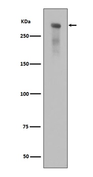
Western blot analysis of Phospho-DNA PKcs (Ser2056) expression in alkaline treated Jurkat cell lysate.
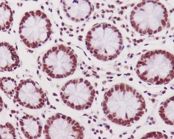
Immunohistochemical analysis of paraffin-embedded human colon tissue using anti-Phospho-DNA PKcs (S2056) antibody. The section was pre-treated using heat mediated antigen retrieval with Tris-EDTA buffer (pH 9.0) for 20 minutes.The tissues were blocked in 5% BSA for 30 minutes at room temperature, washed with ddH2O and PBS, and then probed with the primary antibody (1/200) for 30 minutes at room temperature. The detection was performed using an HRP conjugated compact polymer system. DAB was used as the chromogen. Tissues were counterstained with hematoxylin and mounted with DPX.
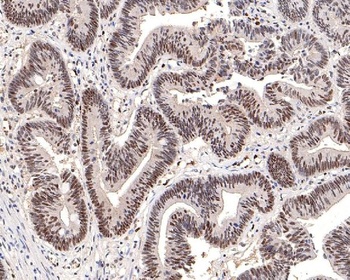
Immunohistochemical analysis of paraffin-embedded human colon carcinoma tissue using anti-Phospho-DNA PKcs (S2056) antibody. The section was pre-treated using heat mediated antigen retrieval with Tris-EDTA buffer (pH 9.0) for 20 minutes.The tissues were blocked in 5% BSA for 30 minutes at room temperature, washed with ddH2O and PBS, and then probed with the primary antibody (1/200) for 30 minutes at room temperature. The detection was performed using an HRP conjugated compact polymer system. DAB was used as the chromogen. Tissues were counterstained with hematoxylin and mounted with DPX.
Submit a review
Filter by Rating

- 5 stars

- 4 stars
- 3 stars
- 2 stars
- 1 stars
