You have no items in your shopping cart.
Cart summary
Item 1 of 5
Item 1 of 5
PHB Antibody
Catalog Number: orb1264666

| Catalog Number | orb1264666 |
|---|---|
| Category | Antibodies |
| Description | PHB Antibody |
| Species/Host | Rabbit |
| Clonality | Polyclonal |
| Tested applications | FC, IF, IHC-P, WB |
| Predicted Reactivity | Bovine, Gallus |
| Reactivity | Human |
| Isotype | Rabbit Ig |
| Immunogen | This PHB antibody is generated from rabbits immunized with a KLH conjugated synthetic peptide between 89-117 amino acids from the Central region of human PHB. |
| Concentration | batch dependent |
| Dilution range | For IHC-P starting dilution is: 1:10~50For WB starting dilution is: 1:1000For IF starting dilution is: 1:10~50For FACS starting dilution is: 1:10~50 |
| Form/Appearance | Liquid |
| Conjugation | Unconjugated |
| MW | 30 kDa |
| Target | PHB |
| UniProt ID | P35232 |
| NCBI | P35232 |
| Storage | Store at 4°C for three months and -20°C, stable for up to one year. As with all antibodies care should be taken to avoid repeated freeze thaw cycles. Antibodies should not be exposed to prolonged high temperatures. |
| Buffer/Preservatives | Supplied in PBS with 0.09% (W/V) sodium azide. |
| Alternative names | Prohibitin, PHB Read more... |
| Note | For research use only |
| Application notes | For IHC-P starting dilution is: 1:10~50For WB starting dilution is: 1:1000For IF starting dilution is: 1:10~50For FACS starting dilution is: 1:10~50 |
| Expiration Date | 12 months from date of receipt. |

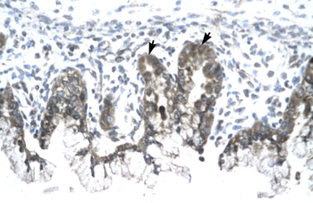
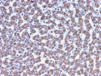
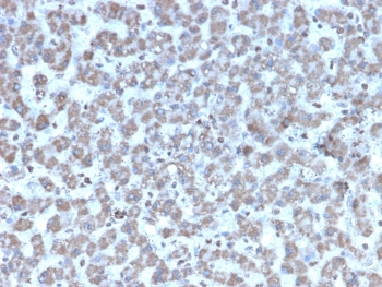
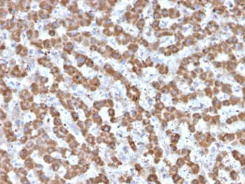
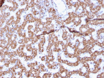
Formalin-fixed and paraffin-embedded human hepatocarcinoma tissue reacted with PHB antibody, which was peroxidase-conjugated to the secondary antibody, followed by DAB staining.

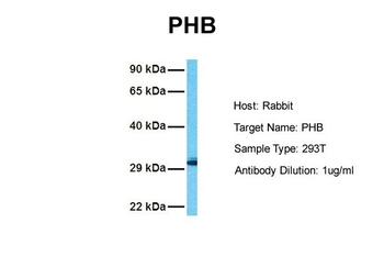
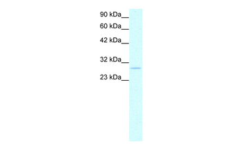
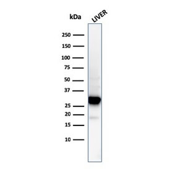
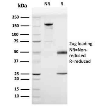
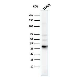
Western blot analysis of PHB Antibody in Hela cell line lysates (35 ug/lane)

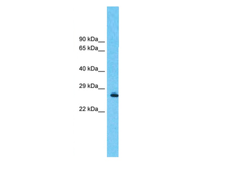
Western blot analysis of PHB using rabbit polyclonal PHB Antibody using 293 cell lysates (2 ug/lane) either nontransfected (Lane 1) or transiently transfected (Lane 2) with the PHB gene.

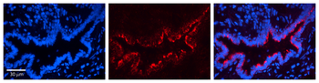
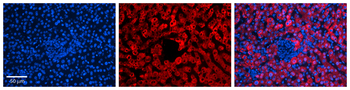
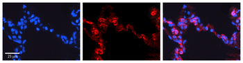
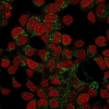
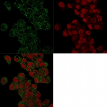
Confocal immunofluorescent analysis of PHB Antibody with Hela cell followed by Alexa Fluor 488-conjugated goat anti-rabbit lgG (green).Actin filaments have been labeled with Alexa Fluor 555 phalloidin (red).

Flow cytometric analysis of Hela cells (right histogram) compared to a negative control cell (left histogram). FITC-conjugated goat-anti-rabbit secondary antibodies were used for the analysis.
- Item 1 of 11
Prohibitin/PHB Antibody [orb745921]
ELISA, FC, IHC, WB
Human, Mouse, Rat
Rabbit
Polyclonal
Unconjugated
10 μg, 100 μg - Item 1 of 9
PHB antibody [orb574394]
IHC, WB
Animal, Bovine, Canine, Guinea pig, Human, Mouse, Rabbit, Rat, Zebrafish
Animal, Bovine, Canine, Guinea pig, Human, Mouse, Rat, Zebrafish
Rabbit
Polyclonal
Unconjugated
100 μl - Item 1 of 5
- Item 1 of 6
- Item 1 of 6


Submit a review
Filter by Rating

- 5 stars

- 4 stars
- 3 stars
- 2 stars
- 1 stars
