You have no items in your shopping cart.
Cart summary
Item 1 of 2
Item 1 of 2
Perforin Antibody / PRF1
Catalog Number: orb1826265

| Catalog Number | orb1826265 |
|---|---|
| Category | Antibodies |
| Description | Perforin is a pore-forming protein that leads to osmotic lysis of the target cells and subsequently enables granzymes to enter the target cells and activate apoptosis. Perforin has structural and functional similarities to complement component 9 (C9). Like C9, this protein creates transmembrane tubules and is capable of lysing non-specifically a variety of target cells. It is one of the main cytolytic proteins of cytolytic granules, and is known to be a key effector molecule for T-cell- and natural killer-cell-mediated cytolysis. Defects in this gene cause familial hemophagocytic lymphohistiocytosis type 2 (HPLH2), a rare and lethal autosomal recessive disorder of early childhood. The expression of perforin is reportedly upregulated in activated CD8+ T-cells, natural killer cells and some CD4+ T-cells. |
| Species/Host | Mouse |
| Clonality | Recombinant |
| Clone Number | rPRF1/8058 |
| Tested applications | IHC-P, WB |
| Reactivity | Human |
| Isotype | Mouse IgG1, kappa |
| Immunogen | A recombinant human Perforin-1 protein fragment (within amino acids 355-555) was used as the immunogen for the PRF1 antibody. |
| Dilution range | Western blot: 1-2ug/ml,Immunohistochemistry (FFPE): 1-2ug/ml for 30 minutes at RT |
| Conjugation | Unconjugated |
| Formula | 0.2 mg/ml in 1X PBS with 0.1 mg/ml BSA (US sourced), 0.05% sodium azide |
| Hazard Information | This PRF1 antibody is available for research use only. |
| UniProt ID | P14222 |
| Storage | Aliquot the PRF1 antibody and store frozen at -20°C or colder. Avoid repeated freeze-thaw cycles. |
| Note | For research use only |
| Expiration Date | 12 months from date of receipt. |
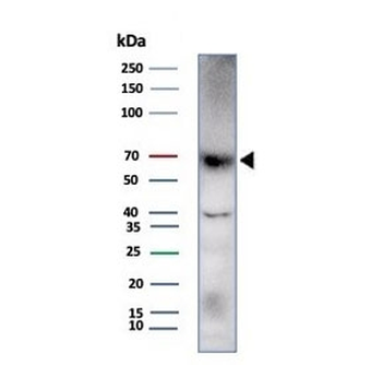
Western blot testing of human HDLM-2 cell lysate with Perforin-1 antibody (clone rPRF1/8058). Predicted molecular weight ~61 kDa.

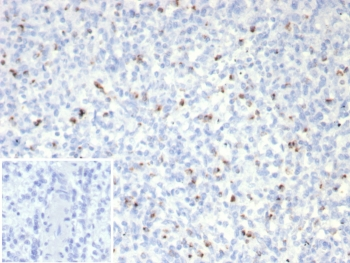
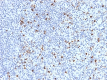
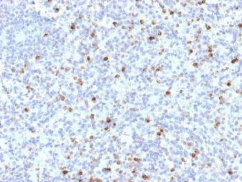
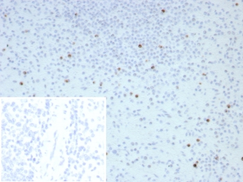
IHC staining of FFPE human spleen tissue with Perforin-1 antibody (clone rPRF1/8058). Inset: PBS used in place of primary Ab (secondary Ab negative control). HIER: boil tissue sections in pH 9 10mM Tris with 1mM EDTA for 20 min and allow to cool before testing.
- Item 1 of 2
- Item 1 of 2
- Item 1 of 2
- Item 1 of 2
- Item 1 of 1


Submit a review
Filter by Rating

- 5 stars

- 4 stars
- 3 stars
- 2 stars
- 1 stars
