You have no items in your shopping cart.
Cart summary
Item 1 of 8
Item 1 of 8
PDCD1 Antibody
Catalog Number: orb1239763

| Catalog Number | orb1239763 |
|---|---|
| Category | Antibodies |
| Description | PDCD1 Antibody |
| Species/Host | Mouse |
| Clonality | Monoclonal |
| Clone Number | 7H6 |
| Tested applications | ELISA, FC, ICC, IF, IHC-P, WB |
| Reactivity | Human |
| Isotype | IgG1 |
| Immunogen | PD-1 antibody was raised against the extracellular domain of human PD-1. |
| Concentration | 1 mg/mL |
| Dilution range | PD-1 antibody can be used for detection of PD-1 by Western blot at 1 μg/mL. Antibody can also be used for immunohistochemistry starting at 5 μg/mL. For immunofluorescence start at 20 μg/mL.Antibody validated: Western Blot in human samples; Immunohistochemistry in human samples; Immunocytochemistry in human samples; Immunofluorescence in human samples and Flow Cytometry in human samples. All other applications and species not yet tested. |
| Form/Appearance | Liquid |
| Conjugation | Unconjugated |
| MW | Predicted: 32 kDa Observed: 38 kDa |
| Target | PDCD1 |
| UniProt ID | Q15116 |
| NCBI | NP_005009 |
| Storage | PD-1 antibody can be stored at 4°C for three months and -20°C, stable for up to one year. As with all antibodies care should be taken to avoid repeated freeze thaw cycles. Antibodies should not be exposed to prolonged high temperatures. |
| Buffer/Preservatives | PD-1 Antibody is supplied in PBS containing 0.02% sodium azide and 50% glycerol. |
| Alternative names | PD-1 Antibody: PD1, PD-1, CD279, SLEB2, hPD-1, hPD Read more... |
| Note | For research use only |
| Application notes | PD-1 antibody can be used for detection of PD-1 by Western blot at 1 μg/mL. Antibody can also be used for immunohistochemistry starting at 5 μg/mL. For immunofluorescence start at 20 μg/mL.Antibody validated: Western Blot in human samples; Immunohistochemistry in human samples; Immunocytochemistry in human samples; Immunofluorescence in human samples and Flow Cytometry in human samples. All other applications and species not yet tested. |
| Expiration Date | 12 months from date of receipt. |

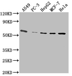
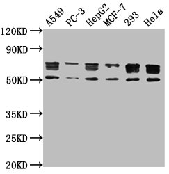
Western blot analysis of PD-1 in transfected 293 cell lysate with PD-1 antibody at 1 μg/mL.

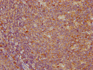
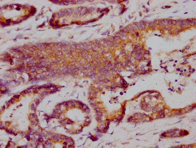
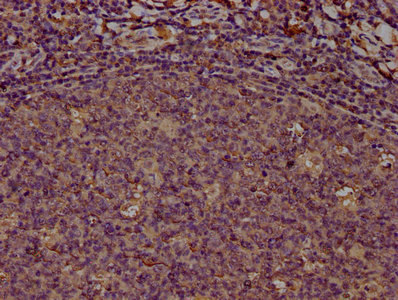
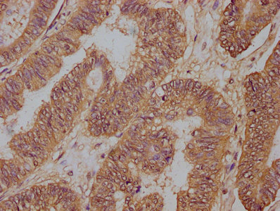
Immunohistochemistry of PD-1 in (A) human tonsil tissue, (B) human lymph node tissue, and (C) human spleen tissue with PD-1 antibody at 5 μg/mL. (D) Immunohistochemistry in human tonsil tissue with control mouse IgG staining at 5 μg/mL.

Immunohistochemistry of PD-1 in (A) human breast cancer tissue and (B) human normal breast tissue with PD-1 antibody at 5 μg/mL.

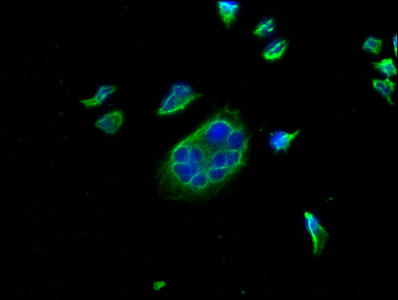
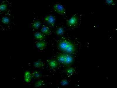
Immunocytochemistry of PD-1 in transfected 293 cells with PD-1 antibody at 5 μg/mL. Lower left: Immunocytochemistry in transfected 293 cells with control mouse IgG antibody at 5 μg/mL.

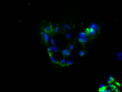
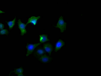
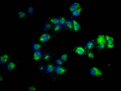
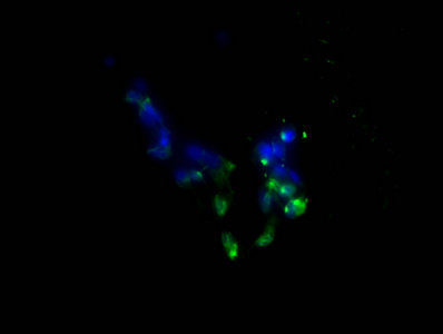
Immunofluorescence of PD-1 in transfected 293 cells with PD-1 antibody at 5 μg/mL. Lower left: Immunofluorescence in transfected 293 cells with control mouse IgG antibody at 5 μg/mL. Red: PD1 Antibody [7H6] (orb1239763) Blue: DAPI staining

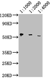
Western blot analysis of PD-1 in overexpressing 293 cells using orb1239745, orb1239769, and orb1239763 antibody at 1, 0.5, and 0.25 μg/ml, respectively.

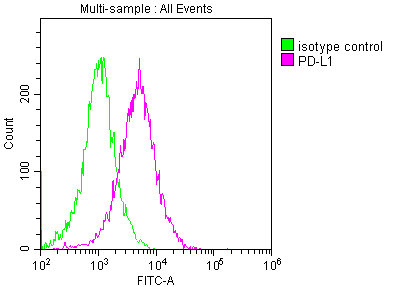
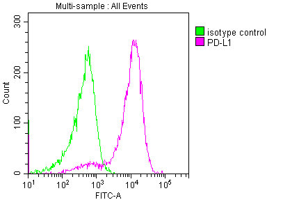
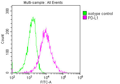
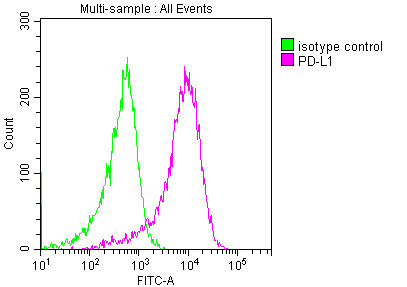
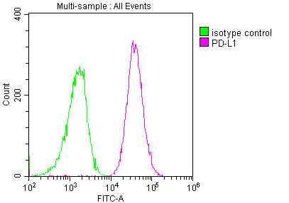
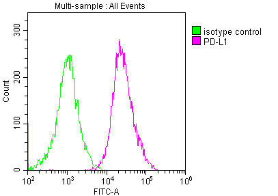
Flow cytometry analysis of PD-1 overexpressing 293 cells using orb1239763 at 1 μg/ml. Blue: untransfected cells, Yellow: PD-1 transfected cells.

Titration curve analysis of PD-1 mAbs to detect recombinant PD-1 in ELISA with orb1239745, orb1239769, orb1239763, orb1239738, and orb1239734 abs at decreasing concentrations.
- Item 1 of 10
- Item 1 of 9
- Item 1 of 9
- Item 1 of 8
PDCD1 Antibody [orb1239732]
ELISA, IF, IHC-P, WB
Human, Mouse, Rat
Rabbit
Polyclonal
Unconjugated
0.1 mg, 0.02 mg - Item 1 of 7


Submit a review
Filter by Rating

- 5 stars

- 4 stars
- 3 stars
- 2 stars
- 1 stars
