You have no items in your shopping cart.
Cart summary
Item 1 of 7
Item 1 of 7
PD L1 Monoclonal Antibody (PDL1)
Catalog Number: orb1787836

| Catalog Number | orb1787836 |
|---|---|
| Category | Antibodies |
| Description | Purified Mouse Monoclonal Antibody (Mab) |
| Species/Host | Mouse |
| Clonality | Monoclonal |
| Tested applications | FC, IHC-P, WB |
| Reactivity | Human |
| Isotype | IgG1 |
| Dilution range | WB: 1:500-1:1000, IHC-P: 1:100-1:600, IHC-P: 1:100-1:600, IHC-P-Leica: 1:100-1:600, IHC-P: 1:100-1:600, IHC-P: 1:100-1:600, IHC-P: 1:100-1:600 |
| Form/Appearance | Purified monoclonal antibody supplied in PBS with 0.09% (W/V) sodium azide. This antibody is purified through a protein G column, followed by dialysis against PBS. |
| Conjugation | Unconjugated |
| MW | 33275 Da |
| Target | This PD L1 antibody is generated from a mouse immunized with a KLH conjugated synthetic peptide between 256-290 amino acids from the human region of human PD L1. |
| UniProt ID | Q9NZQ7 |
| Storage | Maintain refrigerated at 2-8°C for up to 2 weeks. For long term storage store at -20°C in small aliquots to prevent freeze-thaw cycles |
| Alternative names | Programmed cell death 1 ligand 1, PD-L1, PDCD1 lig Read more... |
| Note | For research use only |
| Expiration Date | 12 months from date of receipt. |

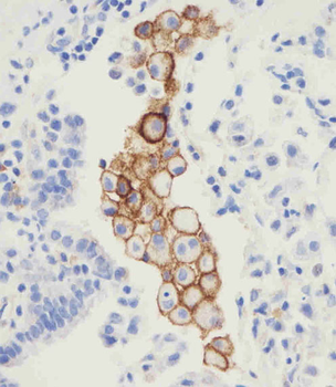
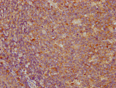
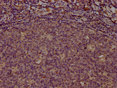
Immunohistochemical analysis of PDL-1 in human tonsil tissue sections (IHC-P - paraformaldehyde-fixed, paraffin-embedded sections) by Dako test. Tissue was fixed with formaldehyde; antigen retrieval was by heat mediation with a EDTA buffer (pH9.0). Samples were incubated with primary antibody (0.85µg/ml) for 1 hours at room temperature. A undiluted biotinylated goat polyvalent antibody was used as the secondary antibody.
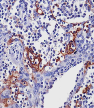
Immunohistochemical analysis of PDL-1 in human tonsil tissue sections (IHC-P - paraformaldehyde-fixed, paraffin-embedded sections) by abgent test. Tissue was fixed with formaldehyde; antigen retrieval was by heat mediation with a EDTA buffer (pH9.0). Samples were incubated with primary antibody (0.85µg/ml) for 1 hours at room temperature. A undiluted biotinylated goat polyvalent antibody was used as the secondary antibody.
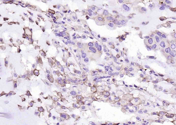
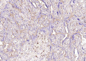
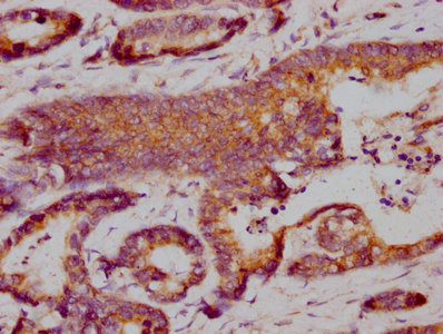
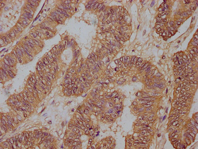
Immunohistochemical analysis of PDL-1 in human non-small cell lung carcinoma sections (IHC-P - paraformaldehyde-fixed, paraffin-embedded sections) by Dako test. Tissue was fixed with formaldehyde; antigen retrieval was by heat mediation with a EDTA buffer (pH9.0). Samples were incubated with primary antibody (0.85µg/ml) for 1 hours at room temperature. A undiluted biotinylated goat polyvalent antibody was used as the secondary antibody.
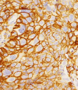
Immunohistochemical analysis of PDL-1 in human non-small cell lung carcinoma sections (IHC-P - paraformaldehyde-fixed, paraffin-embedded sections) by Leica test. Tissue was fixed with formaldehyde; antigen retrieval was by heat mediation with a EDTA buffer (pH9.0). Samples were incubated with primary antibody (0.85µg/ml) for 1 hours at room temperature. A undiluted biotinylated goat polyvalent antibody was used as the secondary antibody.

Immunohistochemical analysis of PDL-1 in human non-small cell lung carcinoma sections (IHC-P - paraformaldehyde-fixed, paraffin-embedded sections) by abgent test. Tissue was fixed with formaldehyde; antigen retrieval was by heat mediation with a EDTA buffer (pH9.0). Samples were incubated with primary antibody (0.85µg/ml) for 1 hours at room temperature. A undiluted biotinylated goat polyvalent antibody was used as the secondary antibody.
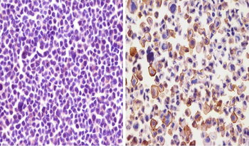
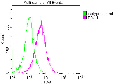
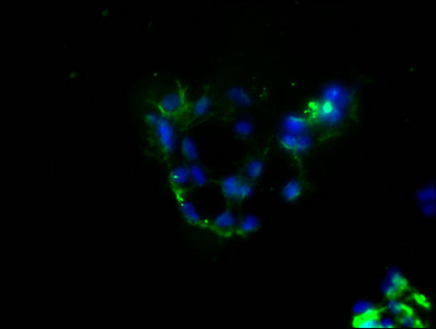
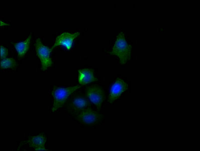
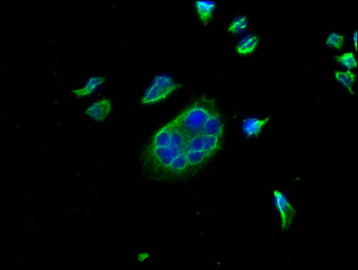
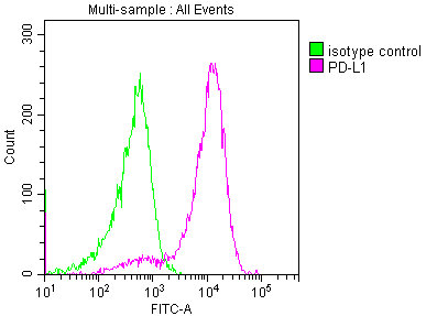
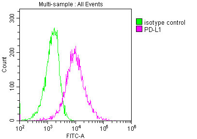
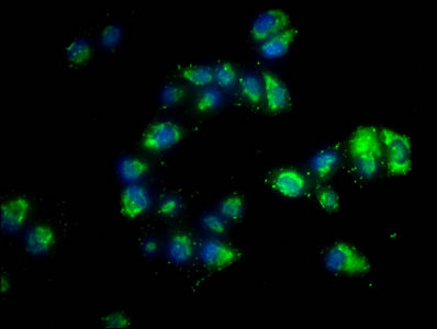
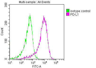
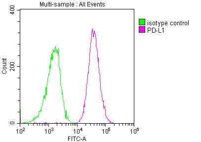
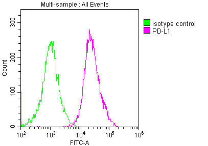
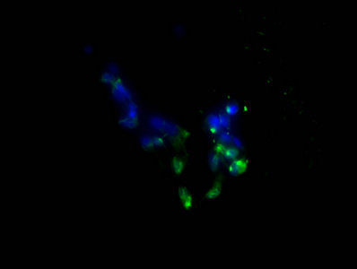
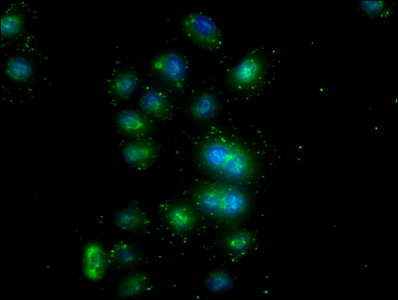
Immunohistochemical analysis of PDL-1 in MCF-7 cell (left) and NCI-H226 (right) cell sections by abgent test.Cell was fixed with formaldehyde and blocked with super block for 10 minutes at room temperature; antigen retrieval was by heat mediation with a EDTA buffer (pH9.0). Samples were incubated with primary antibody (0.85µg/ml) for 1 hours at room temperature. A undiluted biotinylated goat polyvalent antibody was used as the secondary antibody.
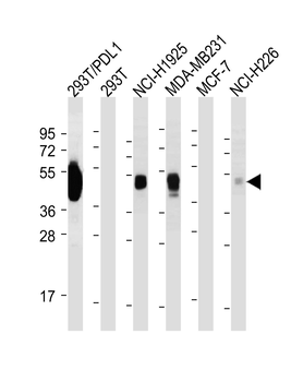
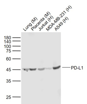
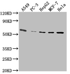
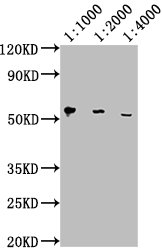
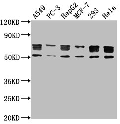
All lanes: Anti-PDL-1 Antibody at 0.5-1µg/ml dilution. Lane 1: 293T/PDL1 whole cell lysate. Lane 2: 293T whole cell lysate. Lane 3: NCI-H1975 whole cell lysate. Lane 4: MDA-MB231 whole cell lysate. Lane 5: MCF-7 whole cell lysate. Lane 6: NCI-H226 whole cell lysate. Lysates/proteins at 30 µg per lane. Secondary Goat Anti-Mouse IgG, (H+L), Peroxidase conjµgated at 1/5000 dilution. Predicted band size: 32 kDa. Blocking/Dilution buffer: 5% NFDM/TBST.
- Item 1 of 10
PD-L1 Monoclonal Antibody [orb688863]
ELISA, FC, IF, IHC, WB
Human
Mouse
Monoclonal
Unconjugated
50 μl, 100 μl - Item 1 of 9
PD-L1 Monoclonal Antibody [orb688864]
ELISA, FC, IF, IHC, WB
Human
Mouse
Monoclonal
Unconjugated
50 μl, 100 μl - Item 1 of 5
- Item 1 of 5
- Item 1 of 4
PD-L1 Recombinant Rabbit Monoclonal Antibody [orb704273]
IF, IHC-Fr, IHC-P, WB
Rat
Human, Mouse
Rabbit
Recombinant
Unconjugated
50 μl, 100 μl, 25 μl
![Anti-PDL1 [YDC 127.1.1]](/images//pub/media/catalog/product/NewWebsite/35/orb613832_1.png)
![Anti-PDL1 [YDC 127.1.1]](/images/pub/media/catalog/product/NewWebsite/35/orb613832_2.png)
![Anti-PDL1 [YDC 127.1.1]](/images/pub/media/catalog/product/NewWebsite/35/orb613832_3.png)
![Anti-PDL1 [YDC 127.1.1]](/images/pub/media/catalog/product/NewWebsite/35/orb613832_4.png)
![Anti-PDL1 [YDC 127.1.1]](/images/pub/media/catalog/product/NewWebsite/35/orb613832_5.png)
![Anti-PDL1 [YDC 127.1.1]](/images//pub/media/catalog/product/NewWebsite/35/orb613833_1.png)
![Anti-PDL1 [YDC 127.1.1]](/images/pub/media/catalog/product/NewWebsite/35/orb613833_2.png)
![Anti-PDL1 [YDC 127.1.1]](/images/pub/media/catalog/product/NewWebsite/35/orb613833_3.png)
![Anti-PDL1 [YDC 127.1.1]](/images/pub/media/catalog/product/NewWebsite/35/orb613833_4.png)
![Anti-PDL1 [YDC 127.1.1]](/images/pub/media/catalog/product/NewWebsite/35/orb613833_5.png)