You have no items in your shopping cart.
Cart summary
Item 1 of 10
Item 1 of 10
PD-L1 Antibody / B7-H1 / CD274
Catalog Number: orb606675

| Catalog Number | orb606675 |
|---|---|
| Category | Antibodies |
| Description | Engagement of CD28 by B7-1 (CD80) or B7-2 (CD86) in the presence of antigen promotes T-cell proliferation, cytokine production, differentiation of effector T-cells and the induction of BCLX, a promoter of T-cell survival. recruitment of CTLA4 by B7-1 or B7-2, on the other hand, may inhibit proliferation and interleukin-2 (IL-2) production. PD-L1 is 290-amino acid type I transmembrane protein, which is 20% and 15% identical to B7-1 and B7-2, respectively, has immunoglobulin V-like and C-like domains and a 30-amino acid cytoplasmic tail. PD-L1 does not bind CD28, cytotoxic T-lymphocyte A4 or ICOS (inducible co-stimulator). IL-2, although produced in small amounts, is required for the effect of PD-L1 co-stimulation. PD-L2 protein contains a signal sequence, IgV- and IgC-like domains, a transmembrane region and a cytoplasmic region. Constitutive expression of PD-L1 and PD-L2 on parenchymal cells of heart, lung and kidney suggests that the PD-1-PD-L system could provide unique negative signaling to help prevent autoimmune diseases. |
| Species/Host | Mouse |
| Clonality | Monoclonal |
| Clone Number | PDL1/2746 |
| Tested applications | ELISA, FACS, IF, IHC-P, WB |
| Reactivity | Human |
| Isotype | Mouse IgG2b, kappa |
| Immunogen | A portion of amino acids 39-191 from the human protein was used as the immunogen for this PD-L1 antibody. |
| Dilution range | ELISA (order BSA/sodium azide-free format for coating),Western blot: 1-2ug/ml,Flow cytometry: 1-2ug/million cells,Immunofluorescence: 1-2ug/ml,Immunohistochemistry (FFPE): 1-2ug/ml for 30 min at RT |
| Purity | Protein G affinity chromatography |
| Conjugation | Unconjugated |
| Formula | 0.2 mg/ml in 1X PBS with 0.1 mg/ml BSA (US sourced) and 0.05% sodium azide |
| Hazard Information | This PD-L1 antibody is available for research use only. |
| UniProt ID | Q9NZQ7 |
| Storage | Store the PD-L1 antibody at 2-8°C (with azide) or aliquot and store at -20°C or colder (without azide). |
| Buffer/Preservatives | 0.2 mg/ml in 1X PBS with 0.1 mg/ml rAlbumin (US sourced) and 0.05% sodium azide |
| Note | For research use only |
| Application notes | Optimal dilution of the PD-L1 antibody should be determined by the researcher. |
| Expiration Date | 12 months from date of receipt. |
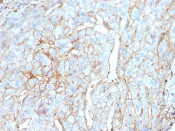
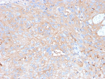
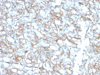
IHC testing of FFPE cervical carcinoma with PD-L1 antibody (clone PDL1/2746). HIER: boil tissue sections in pH9 10mM Tris with 1mM EDTA for 10-20 min followed by cooling at RT for 20 min.
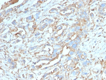
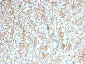
IHC testing of FFPE breast carcinoma with PD-L1 antibody (clone PDL1/2746). HIER: boil tissue sections in pH9 10mM Tris with 1mM EDTA for 10-20 min followed by cooling at RT for 20 min.

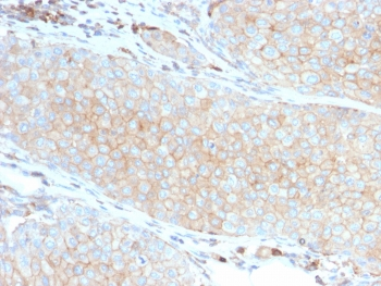
IHC testing of FFPE lung SCC with PD-L1 antibody (clone PDL1/2746). HIER: boil tissue sections in pH9 10mM Tris with 1mM EDTA for 10-20 min followed by cooling at RT for 20 min.

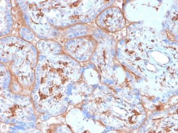
IHC testing of FFPE basal cell carcinoma with PD-L1 antibody (clone PDL1/2746). HIER: boil tissue sections in pH9 10mM Tris with 1mM EDTA for 10-20 min followed by cooling at RT for 20 min.
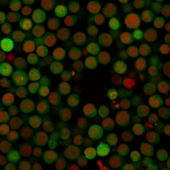
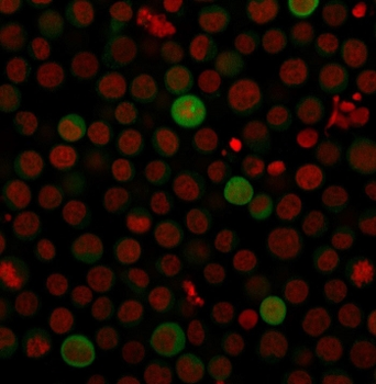
Immunofluorescent staining of human Jurkat cells with PD-L1 antibody (clone PDL1/2746) and a CF488 labeled secondary (green). Nuclei were counterstained with Reddot (red).

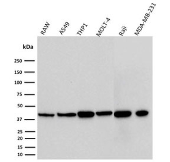
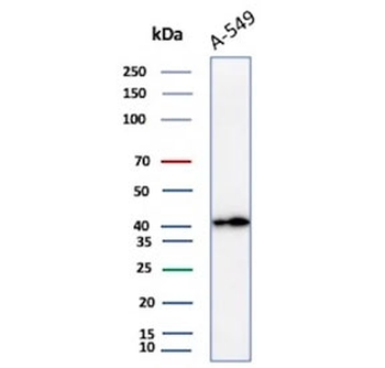
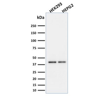
Western blot testing of mouse and human cell lysates with PD-L1 antibody (PDL1/2746). Expected molecular weight ~34 kDa (unmodified), 45-70 kDa (glycosylated).
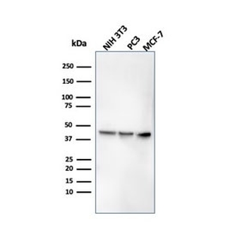
Western blot testing of human cell lysates with PD-L1 antibody (PDL1/2746). Expected molecular weight ~34 kDa (unmodified), 45-70 kDa (glycosylated).
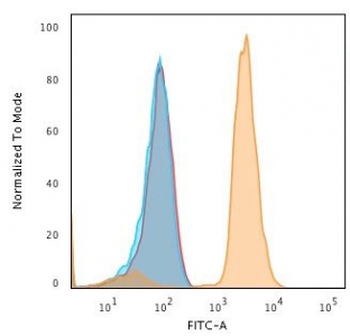
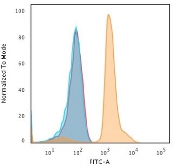
FACS testing of fixed and permeabilized human Jurkat cells with PD-L1 antibody; Blue = cells alone, Red = isotype control, Orange = PD-L1 antibody.

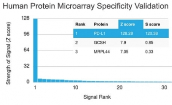
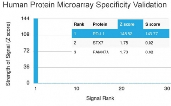
Analysis of HuProt (TM) microarray containing more than 19, 000 full-length human proteins using PD-L1 antibody (clone PDL1/2746). These results demonstrate the foremost specificity of the PDL1/2746 mAb. Z- and S- score: The Z-score represents the strength of a signal that an antibody (in combination with a fluorescently-tagged anti-IgG secondary Ab) produces when binding to a particular protein on the HuProt (TM) array. Z-scores are described in units of standard deviations (SD's) above the mean value of all signals generated on that array. If the targets on the HuProt (TM) are arranged in descending order of the Z-score, the S-score is the difference (also in units of SD's) between the Z-scores. The S-score therefore represents the relative target specificity of an Ab to its intended target.

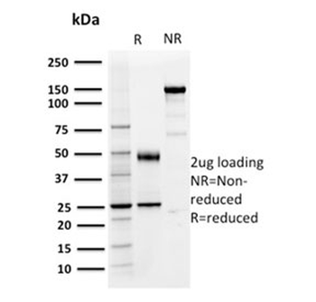
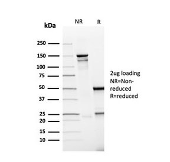
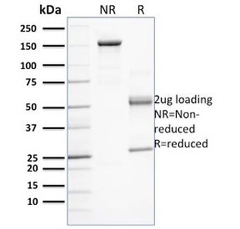
SDS-PAGE analysis of purified, BSA-free PD-L1 antibody (clone PDL1/2746) as confirmation of integrity and purity.
- Item 1 of 10
PD-L1 Antibody / B7-H1 / CD274 [orb2641417]
ELISA, FACS, IF, IHC-P, WB
Human
Mouse
Monoclonal
Unconjugated
100 μg - Item 1 of 5
- Item 1 of 5
- Item 1 of 4
- Item 1 of 4