You have no items in your shopping cart.
Cart summary
Item 1 of 6
Item 1 of 6
PCNA Antibody (C-term)
Catalog Number: orb1927275

| Catalog Number | orb1927275 |
|---|---|
| Category | Antibodies |
| Description | Mouse Monoclonal Antibody (Mab) |
| Species/Host | Mouse |
| Clonality | Monoclonal |
| Clone Number | 1157CT1.1.3 |
| Tested applications | IHC-P, WB |
| Predicted Reactivity | Hamster |
| Reactivity | Human, Mouse, Rat |
| Isotype | IgG1 |
| Dilution range | WB: 1:1000, WB: 1:2000, WB: 1:2000, IHC: 1:2000, IHC-P: 1:25, IHC-P: 1:25 |
| Form/Appearance | Purified monoclonal antibody supplied in PBS with 0.09% (W/V) sodium azide. This antibody is purified through a protein G column, followed by dialysis against PBS. |
| Conjugation | Unconjugated |
| MW | 28769 Da |
| Target | This PCNA antibody is generated from mice immunized with a KLH conjugated synthetic peptide between 236-261 amino acids from the C-terminal region of human PCNA. |
| UniProt ID | P12004 |
| NCBI | NP_002583.1, NP_872590.1 |
| Storage | Maintain refrigerated at 2-8°C for up to 2 weeks. For long term storage store at -20°C in small aliquots to prevent freeze-thaw cycles |
| Alternative names | Proliferating cell nuclear antigen, PCNA, Cyclin, Read more... |
| Note | For research use only |
| Expiration Date | 12 months from date of receipt. |

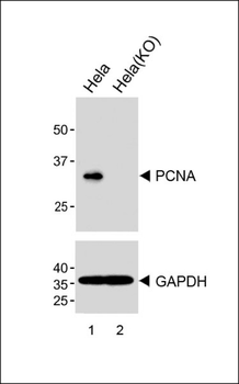
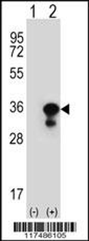
All lanes: Anti-PCNA Antibody (C-term) at 1:1000 dilution (upper) Lane 1: Hela Lane 2: Hela-Knockout Lysates/proteins at 20 µg per lane. Secondary Goat Anti-Mouse IgG, (H+L), Peroxidase conjugated at 1/8000 dilution. Predicted band size: 34 kDa
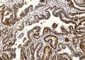
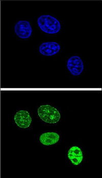
Immunohistochemical analysis of paraffin-embedded Human Ovarian cancer section using Pink1. Diluted at 1:2000 dilution. A undiluted biotinylated goat polyvalent antibody was used as the secondary, followed by DAB staining.

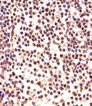
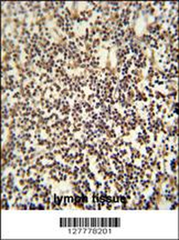
Immunohistochemical analysis of paraffin-embedded H. lymph section using PCNA Antibody (C-term). Diluted at 1:25 dilution. A undiluted biotinylated goat polyvalent antibody was used as the secondary, followed by DAB staining.

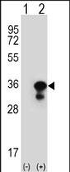
All lanes: Anti-PCNA Antibody (C-term) at 1:2000 dilution + C2C12 whole cell lysate. Lysates/proteins at 20 µg per lane. Secondary Goat Anti-Mouse IgG, (H+L), Peroxidase conjugated at 1/15000 dilution.Observed band size: 34KDa. Blocking/Dilution buffer: 5% NFDM/TBST.
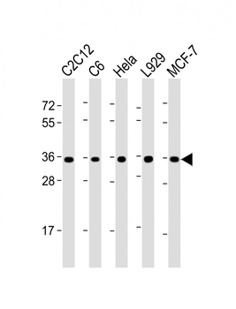
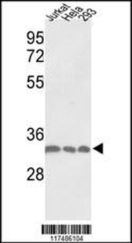
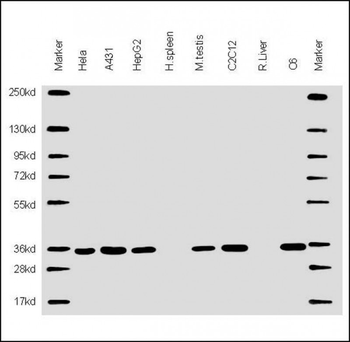
All lanes: Anti-PCNA Antibody (C-term) at 1:2000 dilution. Lane 1: C2C12 whole cell lysate. Lane 2: C6 whole cell lysate. Lane 3: Hela whole cell lysate. Lane 4: L929 whole cell lysate. Lane 5: MCF-7 whole cell lysate. Lysates/proteins at 20 µg per lane. Secondary Goat Anti-mouse IgG, (H+L), Peroxidase conjugated at 1/10000 dilution. Predicted band size: 29 kDa. Blocking/Dilution buffer: 5% NFDM/TBST.
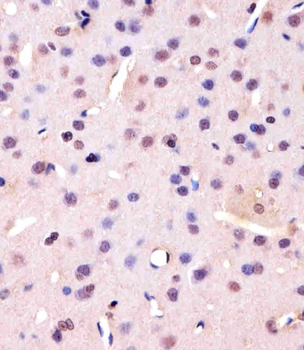
Staining PCNA in Rat brain tissue sections by Immunohistochemistry (IHC-P - paraformaldehyde-fixed, paraffin-embedded sections). Tissue was fixed with formaldehyde and blocked with 3% BSA for 0.5 hour at room temperature; antigen retrieval was by heat mediation with a citrate buffer (pH6). Samples were incubated with primary antibody (1/25) for 1 hours at 37°C. A undiluted biotinylated goat polyvalent antibody was used as the secondary antibody.
- Item 1 of 6
PCNA Antibody (C-term) [orb166911]
IHC-P, WB
Hamster
Human, Mouse, Rat
Mouse
Monoclonal
Unconjugated
80 μl - Item 1 of 6
PCNA Antibody (C-term) [orb1931414]
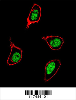
IF, IHC-P, WB
Hamster, Rat
Human
Rabbit
Polyclonal
Unconjugated
400 μl - Item 1 of 6
- Item 1 of 3
PCNA Antibody (C-term) [orb1788295]
IHC, WB
Hamster, Rat
Human, Mouse
Mouse
Monoclonal
Unconjugated
100 μl - Item 1 of 3