You have no items in your shopping cart.
Cart summary
Item 1 of 15
Item 1 of 15
Pan Cytokeratin Rabbit Polyclonal Antibody
Catalog Number: orb10399

| Catalog Number | orb10399 |
|---|---|
| Category | Antibodies |
| Description | Pan Cytokeratin Rabbit Polyclonal Antibody |
| Species/Host | Rabbit |
| Clonality | Polyclonal |
| Tested applications | FC, ICC, IF, IHC-Fr, IHC-P, WB |
| Predicted Reactivity | Canine, Equine, Gallus, Porcine, Rabbit |
| Reactivity | Bovine, Human, Mouse, Rat |
| Isotype | IgG |
| Immunogen | KLH conjugated synthetic peptide derived from human cytokeratins |
| Concentration | 1mg/ml |
| Dilution range | WB=1:500-2000, IHC-P=1:100-2000, IHC-F=1:100-500, ICC/IF=1:100, IF=1:100-500, Flow-Cyt=1μg /test |
| Form/Appearance | Liquid |
| Conjugation | Unconjugated |
| MW | 42-64 kDa |
| UniProt ID | Q2M2I5 |
| RRID | AB_10762614 |
| Storage | Maintain refrigerated at 2-8°C for up to 2 weeks. For long term storage store at -20°C in small aliquots to prevent freeze-thaw cycles. |
| Buffer/Preservatives | 0.01M TBS (pH7.4) with 1% rAlbumin, 0.02% Proclin300 and 50% Glycerol. |
| Alternative names | pan-cytokeratin; pan-CK; pan CK; P-CK; CK-PAN; wid Read more... |
| Note | For research use only |
| Expiration Date | 12 months from date of receipt. |

Blank control (black line): A549. Primary Antibody (green line): Rabbit Anti-Pan Cytokeratin antibody (orb10399), Dilution: 1 ug/Test, Secondary Antibody (white blue line): Goat anti-rabbit IgG-AF488, Dilution: 0.5 ug/Test. Isotype control (orange line): Normal Rabbit IgG, Protocol, The cells were fixed with 4% PFA (10 min at room temperature) and then permeabilized with 90% ice-cold methanol for 20 min at -20°C, The cells were then incubated in 5% BSA to block non-specific protein-protein interactions for 30 min at room temperature. Cells stained with Primary Antibody for 30 min at room temperature. The secondary antibody used for 40 min at room temperature. Acquisition of 20000 events was performed.
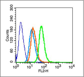
Blank control (blue line): Hela (blue). Primary Antibody (green line): Rabbit Anti-Pan Cytokeratin antibody (orb10399), Dilution: 1 µg/10^6 cells, Isotype Control Antibody (orange line): Rabbit IgG. Secondary Antibody (white blue line): Goat anti-rabbit IgG-PE, Dilution: 1 µg/Test. Protocol, The cells were fixed with 70% methanol (Overnight at 4°C) and then permeabilized with 90% ice-cold methanol for 20 min at -20°C. Cells stained with Primary Antibody for 30 min at room temperature. The cells were then incubated in 1 X PBS/2% BSA/10% goat serum to block non-specific protein-protein interactions followed by the antibody for 15 min at room temperature. The secondary antibody used for 40 min at room temperature. Acquisition of 20000 events was performed.
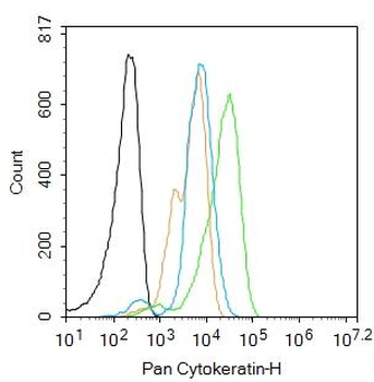
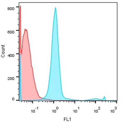
Blank control: Hela. Primary Antibody (green line): Rabbit Anti-Pan Cytokeratin antibody (orb10399), Dilution: 2 ug/Test, Secondary Antibody: Goat anti-rabbit IgG-FITC, Dilution: 0.5 ug/Test. Protocol, The cells were fixed with 4% PFA (10 min at room temperature) and then permeabilized with 0.1% PBST for 20 min at room temperature. The cells were then incubated in 5% BSA to block non-specific protein-protein interactions for 30 min at room temperature. Cells stained with Primary Antibody for 30 min at room temperature. The secondary antibody used for 40 min at room temperature. Acquisition of 20000 events was performed.
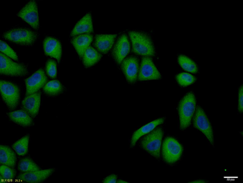
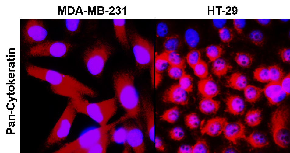
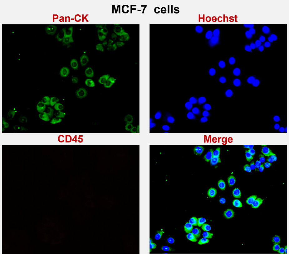
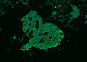
Hela cell, 4% Paraformaldehyde-fixed, Triton X-100 at room temperature for 20 min, Blocking buffer (normal goat serum) at 37°C for 20 min, Antibody incubation with (Pan Cytokeratin) polyclonal Antibody, Unconjugated (orb10399) 1:100, 90 minutes at 37°C, followed by a conjugated Goat Anti-Rabbit IgG antibody at 37°C for 90 minutes, DAPI (blue) was used to stain the cell nuclei.
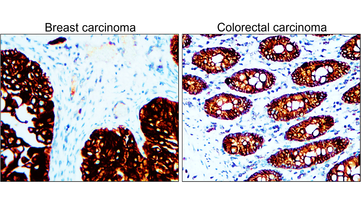
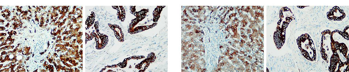
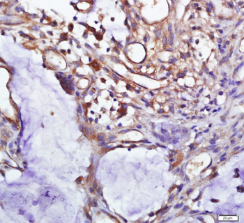
Paraformaldehyde-fixed, paraffin embedded (human breast carcinoma), Antigen retrieval by boiling in sodium citrate buffer (pH6.0) for 15 min, Block endogenous peroxidase by 3% hydrogen peroxide for 20 minutes, Blocking buffer (normal goat serum) at 37°C for 30 min, Antibody incubation with (Pan Cytokeratin) Polyclonal Antibody, Unconjugated (orb10399) at 1:200 overnight at 4°C, followed by operating according to SP Kit (Rabbit) instructionsand DAB staining.
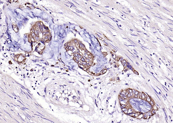
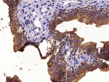
Paraformaldehyde-fixed, paraffin embedded (human cervical cancer), Antigen retrieval by boiling in sodium citrate buffer (pH6.0) for 15 min, Block endogenous peroxidase by 3% hydrogen peroxide for 20 minutes, Blocking buffer (normal goat serum) at 37°C for 30 min, Antibody incubation with (Pan Cytokeratin) Polyclonal Antibody, Unconjugated (orb10399) at 1:200 overnight at 4°C, followed by operating according to SP Kit (Rabbit) instructionsand DAB staining.
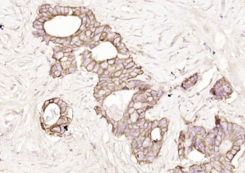
Paraformaldehyde-fixed, paraffin embedded (human cervical carcinoma), Antigen retrieval by boiling in sodium citrate buffer (pH6.0) for 15 min, Block endogenous peroxidase by 3% hydrogen peroxide for 20 minutes, Blocking buffer (normal goat serum) at 37°C for 30 min, Antibody incubation with (Pan Cytokeratin) Polyclonal Antibody, Unconjugated (orb10399) at 1:200 overnight at 4°C, followed by operating according to SP Kit (Rabbit) instructionsand DAB staining.
Paraformaldehyde-fixed, paraffin embedded (human colon carcinoma), Antigen retrieval by boiling in sodium citrate buffer (pH6.0) for 15 min, Blocking buffer (normal goat serum) at 37°C for 30 min, Antibody incubation with (Pan Cytokeratin) Polyclonal Antibody, Unconjugated (orb10399) at 1:200 overnight at 4°C, followed by a conjugated Goat Anti-Rabbit IgG antibody for 90 minutes, and DAPI for nuclei staining.
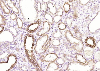
Paraformaldehyde-fixed, paraffin embedded (Human kidney), Antigen retrieval by boiling in sodium citrate buffer (pH6.0) for 15 min, Block endogenous peroxidase by 3% hydrogen peroxide for 20 minutes, Blocking buffer (normal goat serum) at 37°C for 30 min, Antibody incubation with (Pan Cytokeratin) Polyclonal Antibody, Unconjugated (orb10399) at 1:200 overnight at 4°C, followed by operating according to SP Kit (Rabbit) instructionsand DAB staining.
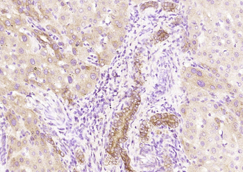
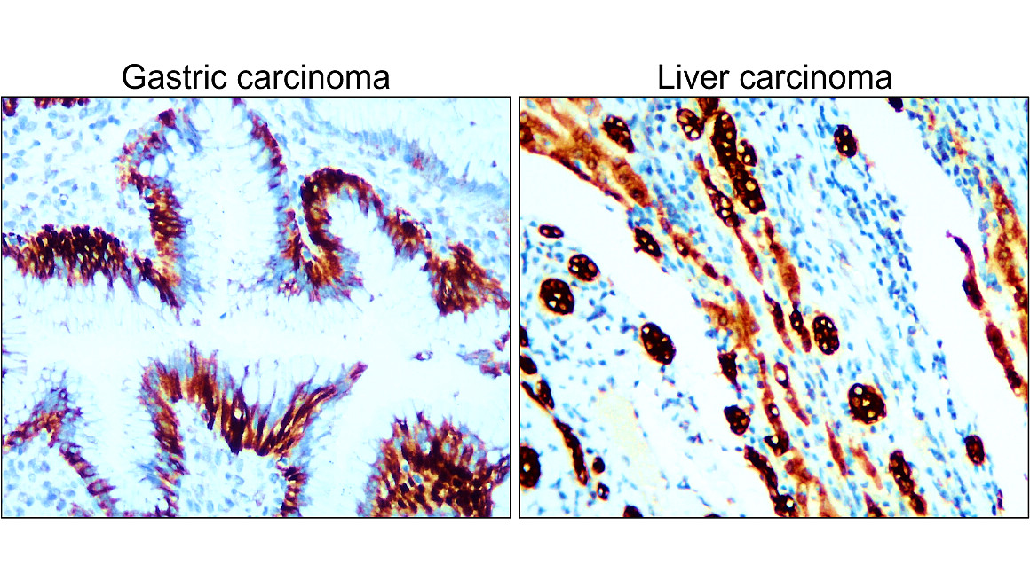
Paraformaldehyde-fixed, paraffin embedded (human liver), Antigen retrieval by boiling in sodium citrate buffer (pH6.0) for 15 min, Block endogenous peroxidase by 3% hydrogen peroxide for 20 minutes, Blocking buffer (normal goat serum) at 37°C for 30 min, Antibody incubation with (Pan Cytokeratin) Polyclonal Antibody, Unconjugated (orb10399) at 1:200 overnight at 4°C, followed by operating according to SP Kit (Rabbit) instructionsand DAB staining.
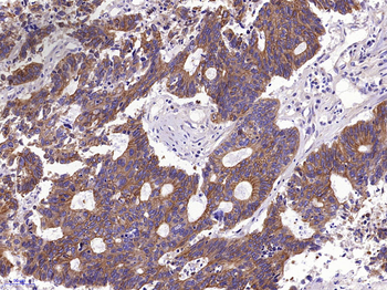
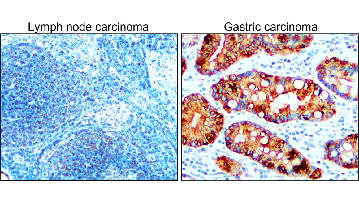
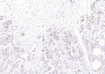
Paraformaldehyde-fixed, paraffin embedded (Human stomach carcinoma), Antigen retrieval by boiling in sodium citrate buffer (pH6.0) for 15 min, Block endogenous peroxidase by 3% hydrogen peroxide for 20 minutes, Blocking buffer (normal goat serum) at 37°C for 30 min, Antibody incubation with (Pan Cytokeratin) Polyclonal Antibody, Unconjugated (orb10399) at 1:400 overnight at 4°C, followed by operating according to SP Kit (Rabbit) instructionsand DAB staining.
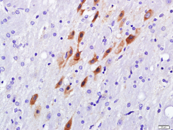
Paraformaldehyde-fixed, paraffin embedded (Rat bladder), Antigen retrieval by boiling in sodium citrate buffer (pH6.0) for 15 min, Block endogenous peroxidase by 3% hydrogen peroxide for 20 minutes, Blocking buffer (normal goat serum) at 37°C for 30 min, Antibody incubation with (Pan Cytokeratin) Polyclonal Antibody, Unconjugated (orb10399) at 1:400 overnight at 4°C, followed by operating according to SP Kit (Rabbit) instructionsand DAB staining.
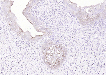
Paraformaldehyde-fixed, paraffin embedded (rat uterus), Antigen retrieval by boiling in sodium citrate buffer (pH6.0) for 15 min, Block endogenous peroxidase by 3% hydrogen peroxide for 20 minutes, Blocking buffer (normal goat serum) at 37°C for 30 min, Antibody incubation with (Pan Cytokeratin) Polyclonal Antibody, Unconjugated (orb10399) at 1:200 overnight at 4°C, followed by operating according to SP Kit (Rabbit) instructionsand DAB staining.
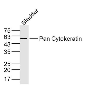
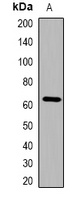
Sample: Bladder (Mouse) Lysate at 40 ug, Primary: Anti-Pan Cytokeratin (orb10399) at 1/300 dilution, Secondary: IRDye800CW Goat Anti-Rabbit IgG at 1/20000 dilution, Predicted band size: 42-64 kD, Observed band size: 60 kD.

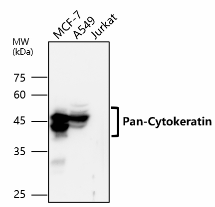
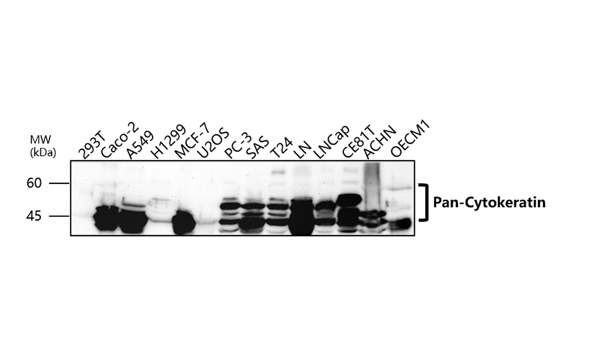
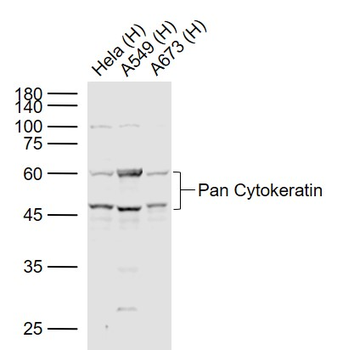
Sample: Lane 1: Hela (Human) Cell Lysate at 30 ug, Lane 2: A549 (Human) Cell Lysate at 30 ug, Lane 3: A673 (Human) Cell Lysate at 30 ug, Primary: Anti-Pan Cytokeratin (orb10399) at 1/1000 dilution, Secondary: IRDye800CW Goat Anti-Rabbit IgG at 1/20000 dilution, Predicted band size: 42-64 kD, Observed band size: 46, 60 kD.
- Item 1 of 12
- Item 1 of 4
Pan Cytokeratin Rabbit Polyclonal Antibody [orb158093]
ELISA, ICC, IF, IHC-Fr, IHC-P, WB
Bovine, Canine, Equine
Human, Mouse, Rat
Rabbit
Polyclonal
Unconjugated
100 μl, 200 μl, 50 μl - Item 1 of 1
Anti-Cytokeratin-pan Antibody [orb382594]
WB
Human, Mouse, Rat
Rabbit
Polyclonal
Unconjugated
200 μl, 100 μl, 30 μl 
Pan Cytokeratin Rabbit Polyclonal Antibody (FITC) [orb15391]
FC, ICC, IF
Canine, Equine, Gallus, Porcine, Rabbit
Bovine, Human, Mouse, Rat
Rabbit
Polyclonal
FITC
100 μl- Item 1 of 2
Cytokeratin-pan (Acetyl Lys194) rabbit pAb [orb763992]
ELISA, WB
Human, Mouse, Rat
Polyclonal
Unconjugated
100 μl, 50 μl