You have no items in your shopping cart.
Cart summary
Item 1 of 3
Item 1 of 3
Pan Cytokeratin Antibody Cocktail (Acidic + Basic)
Catalog Number: orb639459

| Catalog Number | orb639459 |
|---|---|
| Category | Antibodies |
| Description | Twenty human keratins are resolved with two-dimensional gel electrophoresis into acidic (pI 6.0) subfamilies. This pan keratin antibody cocktail recognizes acidic (Type I or LMW) and basic (Type II or HMW) cytokeratins, which include CK1, CK3-6, CK8, CK10, CK14-16, and CK19. Many studies have shown the usefulness of keratin markers in cancer research and tumor diagnosis. The AE1 + AE3 antibody cocktail is a broad spectrum pan cytokeratin antibody cocktail which differentiates epithelial tumors from non-epithelial tumors e.g. squamous vs. adenocarcinoma of the lung, liver carcinoma, breast cancer, and esophageal cancer. It has been used to characterize the source of various neoplasms and to study the distribution of keratin containing cells in epithelia during normal development and during the development of epithelial neoplasms. It stains cytokeratin present in normal and abnormal human tissues and has shown high sensitivity in the recognition of epithelial cells and carcinomas. |
| Species/Host | Mouse |
| Clonality | Monoclonal |
| Clone Number | AE1 + AE3 |
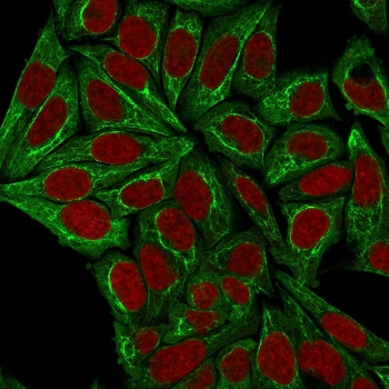
| Tested applications | FACS, IF, IHC, WB |
| Reactivity | Human, Mouse, Rat |
| Isotype | Mouse IgG1, kappa |
| Immunogen | Human epidermal keratin was used as the immunogen for this pan Cytokeratin antibody. |
| Dilution range | FACS: 0.5-1ug/million cells,Immunofluorescence: 1-2ug/ml,Western blot: 0.5-1ug/ml for 2 hours at RT,Immunohistochemistry (FFPE): 0.5-1ug/ml for 30 min at RT (1),Prediluted IHC only format: incubate for 30 min at RT (2) |
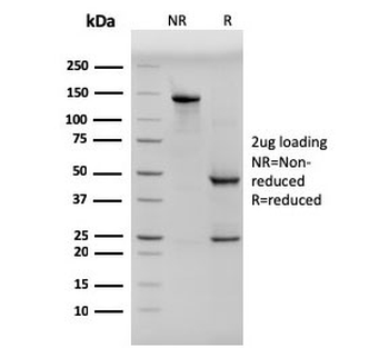
| Purity | Protein G affinity chromatography |
| Conjugation | Unconjugated |
| Formula | 0.2 mg/ml in 1X PBS with 0.1 mg/ml BSA (US sourced) and 0.05% sodium azide |
| Hazard Information | This pan Cytokeratin antibody is available for research use only. |
| Entrez | 1, 3858, 19, 8, 3868, 3848, 15, 3853, 10, 4, 3, 3856, 3866, 3850, 3852, 16, 6, 5, 3880, 3851, 14, 3861 |
| Storage | Store the pan Cytokeratin antibody at 2-8°C (with azide) or aliquot and store at -20°C or colder (without azide). |
| Buffer/Preservatives | 0.2 mg/ml in 1X PBS with 0.1 mg/ml rAlbumin (US sourced) and 0.05% sodium azide |
| Note | For research use only |
| Application notes | The concentration stated for each application is a general starting point. Variations in protocols, secondaries and substrates may require the pan Cytokeratin antibody AE1 + AE3 to be titered up or down for optimal performance.1. Staining of formalin-fixed tissues requires boiling tissue sections in 10mM citrate buffer, pH 6.0, for 10-20 min followed by cooling at RT for 20 minutes.2. The prediluted format is supplied in a dropper bottle and is optimized for use in IHC. After epitope retrieval step (if required), drip mAb solution onto the tissue section and incubate at RT for 30 min. |
| Expiration Date | 12 months from date of receipt. |
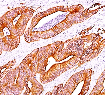
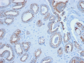
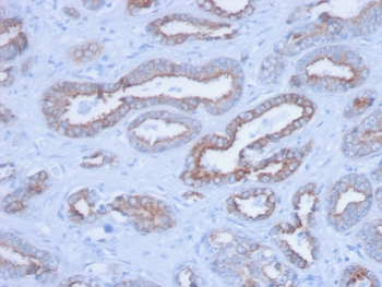
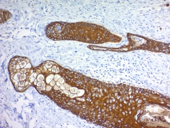
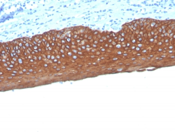
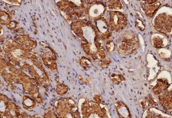
IHC staining of colon carcinoma with pan Cytokeratin antibody cocktail AE1 + AE3.
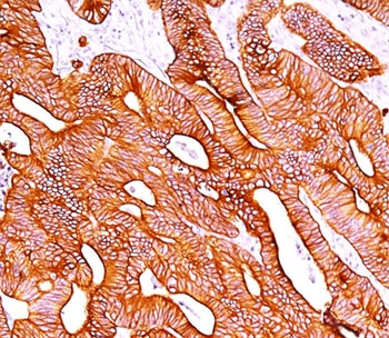
IHC staining of colon carcinoma with pan Cytokeratin antibody cocktail AE1 + AE3.
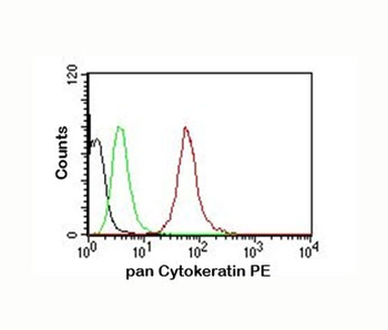
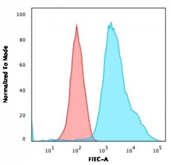
FACS testing of MCF-7 cells: Black = cells alone; Green = isotype control; Red = pan Cytokeratin antibody PE conjugate
- Item 1 of 5
Pan Cytokeratin Antibody Cocktail (Acidic + Basic) [orb640118]
FACS, IF, IHC-P
Human
Mouse
Monoclonal
Unconjugated
20 μg - Item 1 of 5
Pan Cytokeratin Antibody Cocktail (Acidic + Basic) [orb2644183]
FACS, IF, IHC-P
Human
Mouse
Monoclonal
Unconjugated
100 μg - Item 1 of 3
Pan Cytokeratin Antibody Cocktail (Acidic + Basic) [orb1825076]
FACS, IF, IHC-P, WB
Human
Rabbit
Recombinant
Unconjugated
100 μg - Item 1 of 3
Pan Cytokeratin Antibody Cocktail (Acidic + Basic) [orb1825077]
FACS, IF, IHC-P, WB
Human
Rabbit
Recombinant
Unconjugated
20 μg - Item 1 of 3
Pan Cytokeratin Antibody Cocktail (Acidic + Basic) [orb1825078]
FACS, IF, IHC-P, WB
Human
Rabbit
Recombinant
Unconjugated
100 μg