You have no items in your shopping cart.
Cart summary
Item 1 of 2
Item 1 of 2
p53 Antibody / TP53 (N-Terminal Region)
Catalog Number: orb749392

| Catalog Number | orb749392 |
|---|---|
| Category | Antibodies |
| Description | This antibody is specific for a 53kDa protein, which is identified as p53 suppressor gene product. The large number of prolines in its amino acid sequence causes p53 to migrate slowly in SDS-PAGE, resulting in the amino acid content-estimated 43 kDa protein appearing larger than expected. DO-7 reacts with the mutant as well as the wild form of p53 under denaturing and non-denaturing conditions. It binds to MDM2, SV40 T antigen and human papilloma virus E6 protein. Positive nuclear staining with p53 antibody has been reported to be a negative prognostic factor in breast carcinoma, lung carcinoma, colorectal, and urothelial carcinoma. p53 positivity has also been used to differentiate uterine serous carcinoma from endometrioid carcinoma as well as to detect intra-tubular germ cell neoplasia. |
| Species/Host | Mouse |
| Clonality | Monoclonal |
| Clone Number | DO-7 |
| Tested applications | IHC-P, WB |
| Reactivity | Human |
| Isotype | Mouse IgG2b, kappa |
| Immunogen | Recombinant human wild type p53 expressed in E. coli was used as the immunogen for this antibody. Its epitope maps within the N-terminus portion (aa 20-25) of the p53 oncoprotein. |
| Dilution range | Western blot: 0.5-1.0ug/ml,Immunohistochemistry (FFPE): 0.5-1.0ug/ml for 30 min at RT (1),Prediluted IHC only format: incubate for 30 min at RT (2) |
| Purity | Protein G affinity chromatography |
| Conjugation | Unconjugated |
| Formula | 0.2 mg/ml in 1X PBS with 0.1 mg/ml BSA (US sourced) and 0.05% sodium azide |
| Hazard Information | This p53 antibody is available for research use only. |
| Entrez | 7157 |
| Storage | Store the p53 antibody at 2-8°C (with azide) or aliquot and store at -20°C or colder (without azide). |
| Buffer/Preservatives | 0.2 mg/ml in 1X PBS with 0.1 mg/ml rAlbumin (US sourced) and 0.05% sodium azide |
| Note | For research use only |
| Application notes | The concentration stated for each application is a general starting point. Variations in protocols, secondaries and substrates may require the p53 antibody to be titered up or down for optimal performance.1. Staining of formalin-fixed tissues requires boiling tissue sections in 10mM citrate buffer, pH 6.0, for 10-20 min followed by cooling at RT for 20 minutes.2. The prediluted format is supplied in a dropper bottle and is optimized for use in IHC. After epitope retrieval step (if required), drip mAb solution onto the tissue section and incubate at RT for 30 min. |
| Expiration Date | 12 months from date of receipt. |
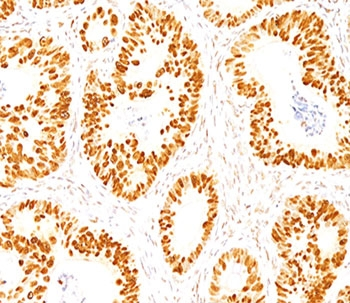
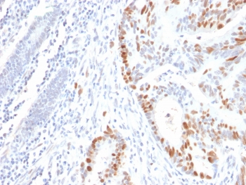
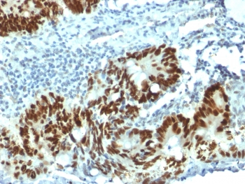
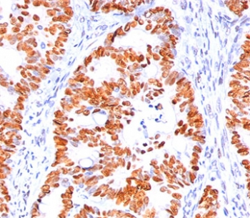
IHC staining of FFPE normal human colon with p53 antibody (clone DO-7).
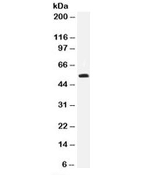
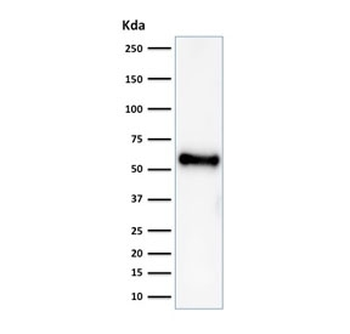
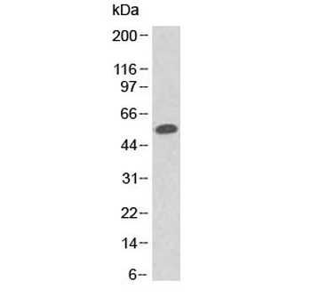
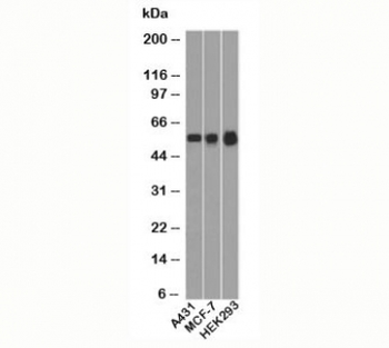
Western blot testing of 293 cell lysate with p53 antibody (clone DO-7).
- Item 1 of 4
p53 Antibody / TP53 N-Terminal Region [orb606470]
IHC-P, WB
Human
Mouse
Recombinant
Unconjugated
100 μg, 20 μg - Item 1 of 3
p53 Antibody / TP53 (N-Terminal Region) [orb402962]
FACS, IF, IHC-P, WB
Human
Mouse
Monoclonal
Unconjugated
100 μg, 20 μg - Item 1 of 2
p53 Antibody / TP53 (N-Terminal Region) [orb749391]
IHC-P, WB
Human
Mouse
Monoclonal
Unconjugated
20 μg, 100 μg
Submit a review
Filter by Rating

- 5 stars

- 4 stars
- 3 stars
- 2 stars
- 1 stars
