You have no items in your shopping cart.
p38 Antibody (FITC)
SKU: orb151880
Description
Images & Validation
−Item 1 of 3
| Tested Applications | ICC, IF, IHC, IP, WB |
|---|---|
| Dilution range | WB (1:1000), ICC/IF (1:100), IP (1:250) |
| Reactivity | Bovine, Canine, Gallus, Guinea pig, Hamster, Human, Monkey, Mouse, Porcine, Rabbit, Rat, Sheep |
| Application Notes |
Key Properties
−| Host | Rabbit |
|---|---|
| Clonality | Polyclonal |
| Immunogen | A 20 residue synthetic peptide based on the human p38 with the cysteine residue added and coupled to KLH |
| Target | p38 |
| Molecular Weight | 43kDa |
| Purification | Peptide Affinity Purified |
Storage & Handling
−| Storage | Conjugated antibodies should be stored according to the product label |
|---|---|
| Buffer/Preservatives | 640.91mM DMSO, 136.36 mM Ethanolamine, 126.89 mM chlorides, 9.09mM phosphates, 9.09mM NaHCO3 |
| Concentration | 1 mg/ml |
| Disclaimer | For research use only |
Alternative Names
−CSAID Binding protein 1, CSBP1, CSBP2, EXIP, MAP kinase MXI2, MAPkinase p38alpha, MAPK14, p38 ALPHA, p38 MAP kinase, p38 mitogen activated protein kinase, RK, SAPK 2A, Stress activated protein kinase 2A
Similar Products
−- Item 1 of 6
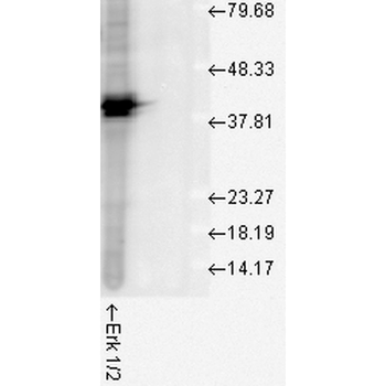
Erk1/2 Antibody (FITC) [orb151497]
FC, ICC, IF, IHC, WB
Bovine, Drosophila, Frog, Gallus, Human, Mouse, Rat, Sheep
Rabbit
Polyclonal
FITC
100 μl - Item 1 of 4
AHA1 Antibody (FITC) [orb147671]
ELISA, ICC, IF, IHC, IP, WB
Human, Mouse, Rat
Rat
Monoclonal
FITC
100 μg - Item 1 of 4
AHA1 Antibody (FITC) [orb147688]
ELISA, ICC, IF, IHC, IP, WB
Human, Mouse, Rat
Rat
Monoclonal
FITC
100 μg - Item 1 of 3
p38 MAPK Antibody (FITC) [orb147365]
AM, ELISA, IHC, IP, WB
Human, Mouse, Rat
Mouse
Monoclonal
FITC
100 μg - Item 1 of 2
AHA1 Antibody (FITC) [orb152016]
ELISA, ICC, IF, IHC, IP, WB
Human, Mouse, Rat
Rabbit
Polyclonal
FITC
100 μl


Quality Guarantee
Explore bioreagents carefree to elevate your research. All our products are rigorously tested for performance. If a product does not perform as described on its datasheet, our scientific support team will provide expert troubleshooting, a prompt replacement, or a refund. For full details, please see our Terms & Conditions and Buying Guide. Contact us at [email protected].
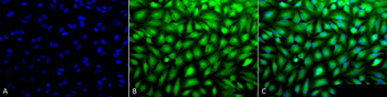
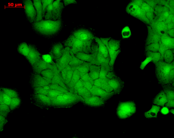
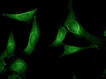
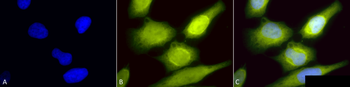
Immunocytochemistry/Immunofluorescence analysis using Rabbit Anti-p38 Polyclonal Antibody. Tissue: Cervical cancer cell line (HeLa). Species: Human. Fixation: 2% Formaldehyde for 20 min at RT. Primary Antibody: Rabbit Anti-p38 Polyclonal Antibody at 1:100 for 12 hours at 4°C. Secondary Antibody: FITC Goat Anti-Rabbit (green) at 1:200 for 2 hours at RT. Counterstain: DAPI (blue) nuclear stain at 1:40000 for 2 hours at RT. Localization: Mitochondrion. Cytoplasm. Nucleus. Magnification: 100x. (A) DAPI (blue) nuclear stain. (B) Anti-p38 Antibody. (C) Composite.
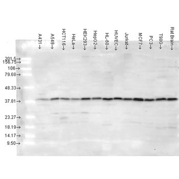
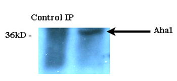
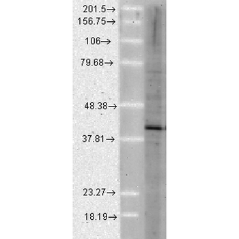
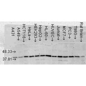
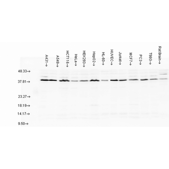
Western blot analysis of Human cancer cell lines showing detection of p38 protein using Rabbit Anti-p38 Polyclonal Antibody. Load: 15 μgprotein. Block: 1.5% BSA for 30 minutes at RT. Primary Antibody: Rabbit Anti-p38 Polyclonal Antibody at 1:4000 for 2 hours at RT. Secondary Antibody: Donkey Anti-Rabbit IgG: HRP for 1 hour at RT.

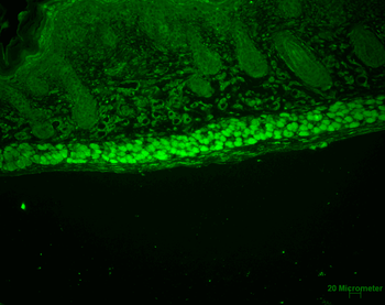
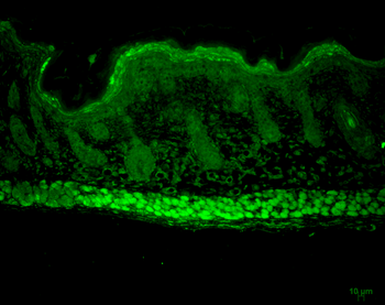
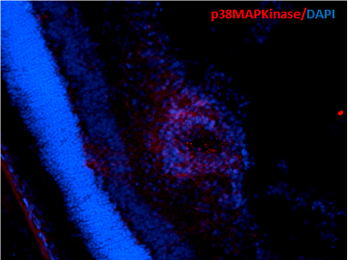
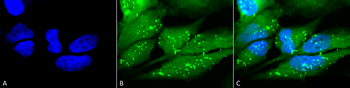
Immunocytochemistry/Immunofluorescence analysis using Rabbit Anti-p38 Polyclonal Antibody. Tissue: Cervical cancer cell line (HeLa). Species: Human. Fixation: 2% Formaldehyde for 20 min at RT. Primary Antibody: Rabbit Anti-p38 Polyclonal Antibody at 1:100 for 12 hours at 4°C. Secondary Antibody: FITC Goat Anti-Rabbit (green) at 1:200 for 2 hours at RT. Counterstain: DAPI (blue) nuclear stain at 1:40000 for 2 hours at RT. Localization: Mitochondrion. Cytoplasm. Nucleus. Magnification: 20x. (A) DAPI (blue) nuclear stain. (B) Anti-p38 Antibody. (C) Composite.
Quick Database Links
UniProt Details
− No UniProt data available
NCBI Gene Details
− No NCBI Gene data available
NCBI Reference Sequences
−Associated Accession Numbers
Curated reference sequences for the gene transcript and protein product| Protein | NP_001306.1 |
|---|
Documents Download
Datasheet
Product Information
Request a Document
Protocol Information
WB
Western Blot (IB, immunoblot)
IHC
Immunohistochemistry
IF
Immunofluorescence
ICC
Immunocytochemistry
IP
Immunoprecipitation
p38 Antibody (FITC) (orb151880)

- 0.0
Based on 0 reviews
Participating in our Biorbyt product reviews program enables you to support fellow scientists by sharing your firsthand experience with our products.
Login to Submit a ReviewAvailable Sizes
Select a size below