You have no items in your shopping cart.
Cart summary
Item 1 of 7
Item 1 of 7
P2RX7 antibody
Catalog Number: orb20291

| Catalog Number | orb20291 |
|---|---|
| Category | Antibodies |
| Description | Goat polyclonal antibody to P2RX7 |
| Species/Host | Goat |
| Clonality | Polyclonal |
| Tested applications | ELISA, FC, IF, IHC, WB |
| Reactivity | Canine, Human, Mouse, Rat |
| Dilution range | ELISA: 1:16000, WB: 1-3 μg/ml, IHC-P: 3.75 μg/ml |
| Conjugation | Unconjugated |
| MW | 68.6 |
| Target | P2RX7 / P2X7 receptor |
| Entrez | 5027 |
| Protein Sequence | YETNKVTRIQSMNY |
| RRID | AB_10753748 |
| Storage | Aliquot and store at -20°C. Minimize freezing and thawing. |
| Buffer/Preservatives | Supplied at 0.5 mg/ml in Tris saline, 0.02% sodium azide, pH 7.3 with 0.5% bovine serum albumin. Aliquot and store at -20°C. Minimize freezing and thawing. |
| Alternative names | anti P2RX7 antibody, anti purinergic receptor P2X, Read more... |
| Note | For research use only |
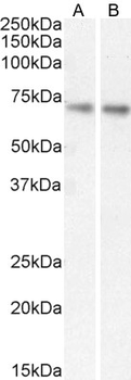
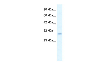
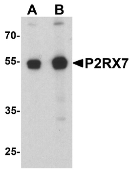
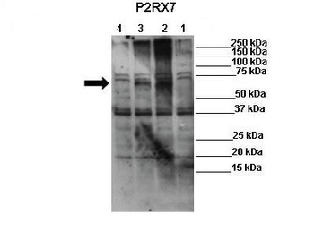
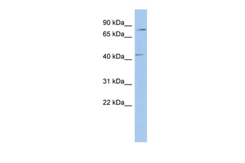
| Application notes | ELISA: Peptide ELISA: antibody detection limit dilution 1:16000.WB: Approx 37kDa and 70kDa band observed in Human Brain (Frontal Cortex) lysates (calculated MW of 68.6kDa according to NP_002553.2). Recommended concentration: 1-3 μg/ml. |
| Expiration Date | 12 months from date of receipt. |
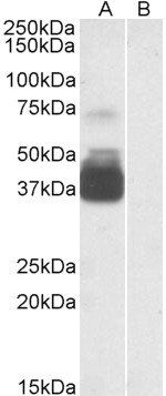
Western blot analysis of Human Brain (Frontal Cortex) lysate using P2RX7 antibody
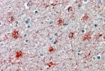
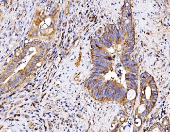
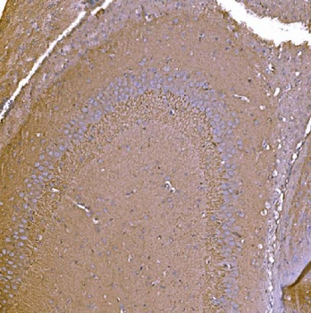
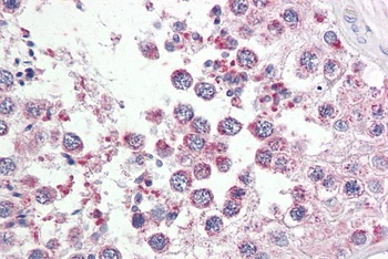
Immunohistochemical staining of Human Brain Cortex using P2RX7 antibody
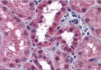
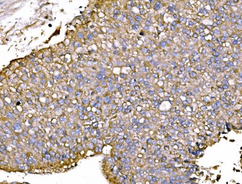
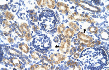
Immunohistochemical staining of Human Kidney using P2RX7 antibody
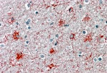
orb20291 (3.75 µg/mL) staining of paraffin embedded Human Cortex. Steamed antigen retrieval with citrate buffer pH 6, AP-staining.
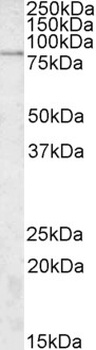
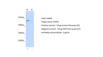
orb20291 (0.3 µg/mL) staining of A431 (A) and HeLa (B) cell lysate (35 µg protein in RIPA buffer). Detected by chemiluminescence.
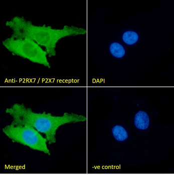
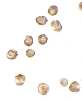
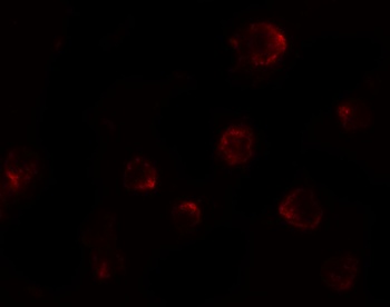
orb20291 Immunofluorescence analysis of paraformaldehyde fixed HeLa cells, permeabilized with 0.15% Triton. Primary incubation 1 hr (10 µg/mL) followed by Alexa Fluor 488 secondary antibody (2 µg/mL), showing cytoplasmic and plasma membrane staining. The nuclear stain is DAPI (blue). Negative control: Unimmunized goat IgG (10 µg/mL) followed by Alexa Fluor 488 secondary antibody (2 µg/mL).
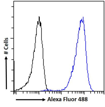
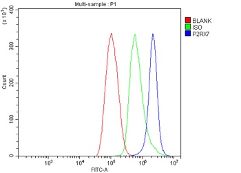
orb20291 Flow cytometric analysis of paraformaldehyde fixed HeLa cells (blue line), permeabilized with 0.5% Triton. Primary incubation 1 hr (10 µg/mL) followed by Alexa Fluor 488 secondary antibody (1 µg/mL). IgG control: Unimmunized goat IgG (black line) followed by Alexa Fluor 488 secondary antibody.
- Item 1 of 5
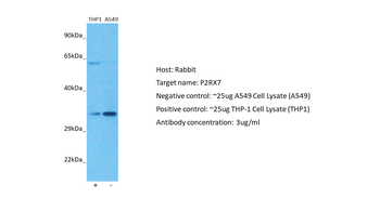
P2X7/P2RX7 Antibody [orb763184]
ELISA, FC, IHC, WB
Human, Mouse, Rat
Rabbit
Polyclonal
Unconjugated
10 μg, 100 μg - Item 1 of 4
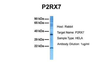
P2RX7 antibody [orb575528]
IHC, WB
Bovine, Canine, Equine, Guinea pig, Mouse, Rabbit, Rat
Human
Rabbit
Polyclonal
Unconjugated
100 μl - Item 1 of 3
P2RX7 Antibody [orb1239302]
ELISA, ICC, IF, WB
Rat
Human, Mouse
Rabbit
Polyclonal
Unconjugated
0.1 mg, 0.02 mg - Item 1 of 3
- Item 1 of 3
P2RX7 antibody [orb575375]
WB
Bovine, Canine, Equine, Guinea pig, Mouse, Porcine, Rabbit, Rat
Human
Rabbit
Polyclonal
Unconjugated
100 μl
Submit a review
Filter by Rating

- 5 stars

- 4 stars
- 3 stars
- 2 stars
- 1 stars
