You have no items in your shopping cart.
Cart summary
Item 1 of 7
Item 1 of 7
NPTX2 Antibody
Catalog Number: orb1239671

| Catalog Number | orb1239671 |
|---|---|
| Category | Antibodies |
| Description | NPTX2 Antibody |
| Species/Host | Rabbit |
| Clonality | Polyclonal |
| Tested applications | ELISA, IF, IHC-P, WB |
| Predicted Reactivity | Guinea pig |
| Reactivity | Human, Mouse, Rat |
| Isotype | IgG |
| Immunogen | Anti-NPTX2 antibody (orb1239671) was raised against a peptide corresponding to 16 amino acids near the center of human NPTX2. The immunogen is located within amino acids 170 - 220 of NPTX2. |
| Concentration | 1 mg/mL |
| Form/Appearance | Liquid |
| Conjugation | Unconjugated |
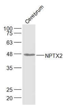
| MW | Predicted: 47kDObserved: 47 kD |
| Target | NPTX2 |
| UniProt ID | P47972 |
| NCBI | NP_002514 |
| Storage | Maintain refrigerated at 2-8°C for up to 2 weeks. For long term storage store at -20°C in small aliquots to prevent freeze-thaw cycles. |
| Buffer/Preservatives | NPTX2 Antibody is supplied in PBS containing 0.02% sodium azide. |
| Alternative names | NPTX2 Antibody: NP2, NARP, NP-II, Neuronal pentrax Read more... |
| Note | For research use only |
| Expiration Date | 12 months from date of receipt. |

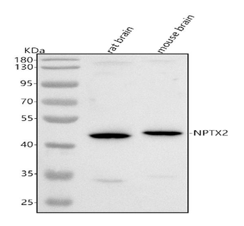
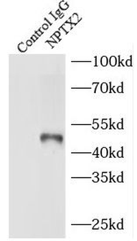
Western Blot Validation in Mouse Brain Tissue Lysate. Loading: 15 µg of lysates per lane. Antibodies: NPTX2 orb1239671 (A: 0.5 µg/mL, B: 1 µg/mL), 1h incubation at RT in 5% NFDM/TBST. Secondary: Goat anti-rabbit IgG HRP conjugate at 1:10000 dilution.

Independent Antibody Validation (IAV) via Protein Expression Profile in Cell Lines. Loading: 15 µg of lysates per lane. Antibodies: NPTX2 orb1239671 (2 µg/mL), NPTX2 orb1271003 (4 µg/mL), and beta-actin orb1240312 (1 µg/mL), 1h incubation at RT in 5% NFDM/TBST. Secondary: Goat anti-rabbit IgG HRP conjugate at 1:10000 dilution.

Western Blot Validation in Mouse Brain Tissue Lysate. Loading: 15 µg of lysates per lane. Antibodies: NPTX2 orb1239671 (1 µg/mL), 1h incubation at RT in 5% NFDM/TBST. Secondary: Goat anti-rabbit IgG HRP conjugate at 1:10000 dilution. A: Absence of blocking peptide B: Presence of blocking peptide.

Immunofluorescence Validation of NPTX2 in Human Brain Tissue. Immunofluorescent analysis of 4% paraformaldehyde-fixed human brain tissue labeling NPTX2 with orb1239671 at 20 µg/mL, followed by goat anti-rabbit IgG secondary antibody at 1/500 dilution (red).

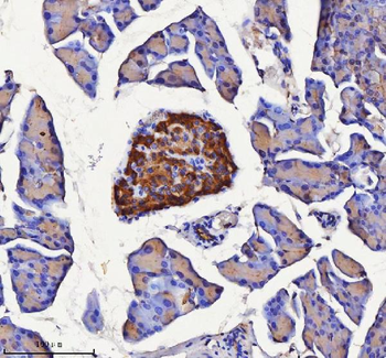
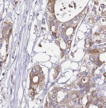
Immunohistochemistry Validation of NPTX2 in Human Brain. Immunohistochemical analysis of paraffin-embedded human brain using anti-NPTX2 antibody (orb1239671) at 5 µg/ml. Tissue was fixed with formaldehyde and blocked with 10% serum for 1 h at RT; antigen retrieval was by heat mediation with a citrate buffer (pH6). Samples were incubated with primary antibody overnight at 4 °C. A goat anti-rabbit IgG H&L (HRP) at 1/250 was used as secondary. Counter stained with Hematoxylin.

Immunofluorescence Validation of NPTX2 in Mouse Brain Tissue. Immunofluorescent analysis of 4% paraformaldehyde-fixed mouse brain issue labeling NPTX2 with orb1239671 at 20 µg/mL, followed by goat anti-rabbit IgG secondary antibody at 1/500 dilution (red) and DAPI staining (blue).

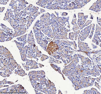
Immunohistochemistry Validation of NPTX2 in Mouse Brain Tissue. Immunohistochemical analysis of paraffin-embedded mouse brain issue using anti-NPTX2 antibody (orb1239671) at 5 µg/ml. Tissue was fixed with formaldehyde and blocked with 10% serum for 1 h at RT; antigen retrieval was by heat mediation with a citrate buffer (pH6). Samples were incubated with primary antibody overnight at 4°C. A goat anti-rabbit IgG H&L (HRP) at 1/250 was used as secondary. Counter stained with Hematoxylin.
- Item 1 of 5
Anti-NPTX2 Antibody [orb1939837]
ELISA, IHC, WB
Human, Mouse, Rat
Rabbit
Polyclonal
Unconjugated
10 μg, 100 μg - Item 1 of 3
NPTX2 Antibody [orb629386]
ELISA, IHC, IP, WB
Human, Mouse, Rat
Rabbit
Polyclonal
Unconjugated
100 μg, 50 μg - Item 1 of 1
- Item 1 of 2
NPTX2 Rabbit Polyclonal Antibody [orb2841]
IF, IHC-Fr, IHC-P, WB
Bovine, Canine, Human, Rat, Sheep
Mouse
Rabbit
Polyclonal
Unconjugated
50 μl, 100 μl, 200 μl - Item 1 of 2