You have no items in your shopping cart.
Cart summary
Item 1 of 5
Item 1 of 5
NOTCH1 antibody
Catalog Number: orb750485

| Catalog Number | orb750485 |
|---|---|
| Category | Antibodies |
| Description | NOTCH1 antibody |
| Species/Host | Rabbit |
| Clonality | Polyclonal |
| Tested applications | DOT, ELISA, IF, IHC, IP, WB |
| Reactivity | Human, Mouse |
| Isotype | Antiserum |
| Immunogen | This whole rabbit serum was prepared by repeated immunizations with a synthetic peptide corresponding to amino acid residues of human Notch 1 located near the N-terminal sequence of the cleaved N intracellular domain (NICD). |
| Concentration | 80 mg/mL |
| Dilution range | ELISA: 1:20,000 - 1:60,000, IHC: 1:200, IF: User Optimized, IP: User Optimized, WB: 1:500- 1:2,000 |
| Form/Appearance | Liquid (sterile filtered) |
| Purity | This antiserum is directed against human NOTCH 1. Based on the immunogen sequence, we expect this antibody to react as well with mouse and rat NOTCH 1 (100% sequence homology). This antibody reacts with mouse Notch constructs present in lysates of HEK 293 cells. Only the cleaved intracellular (activated) form (NICD) is detected. No reactivity is detected against mouse N2, N3 or N4. The immunogen epitope is only exposed after gamma secretase cleavage and is not accessible in the uncleaved form. |
| Conjugation | Unconjugated |
| UniProt ID | P46531 |
| NCBI | NP_060087.3 |
| Storage | Store vial at -20° C or below prior to opening. This vial contains a relatively low volume of reagent (25 µL). To minimize loss of volume dilute 1:10 by adding 225 µL of the buffer stated above directly to the vial. Recap, mix thoroughly and briefly centrifuge to collect the volume at the bottom of the vial. Use this intermediate dilution when calculating final dilutions as recommended below. Store the vial at -20°C or below after dilution. Avoid cycles of freezing and thawing. |
| Buffer/Preservatives | 0.1% (w/v) Sodium Azide |
| Alternative names | rabbit anti-notch1 antibody, Neurogenic locus Notc Read more... |
| Note | For research use only |
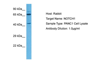
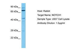
| Application notes | Anti-Notch 1 has been tested by ELISA, dot blot, western blot and immunohistochemistry. An 80 kDa band corresponding to Notch 1 was observed at a 1:500 dilution. Specific conditions for reactivity should be optimized by the end user. |
| Expiration Date | 12 months from date of receipt. |
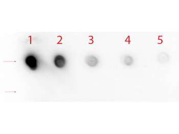
Dot Blot of Rabbit anti-Notch 1 (Cleaved N Terminal) (Human Specific) Antibody. Antigen: Row 1 - Notch 1 Peptide (Cleaved N Terminal) Row 2 - Notch 1 (Intra) Peptide. Load: Lane 1 - 200 ng Lane 2 - 66.67 ng Lane 3 - 22.22 ng Lane 4 - 7.41 ng Lane 5 - 2.47 ng. Primary antibody: Rabbit anti-Notch 1 (Cleaved N Terminal) (Human Specific) Antibody at 1:1000 for 60 min at RT. Secondary antibody: HRP Rabbit Secondary at 1:40000 for 30 min at RT. Block: orb348637 for 1 HR at RT.

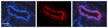
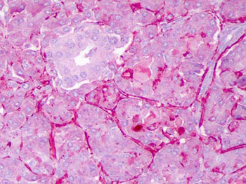
Immunohistochemistry of Rabbit anti-Notch1 antibody. Tissue: Exocrine glands of human pancreas. Fixation: FFPE. Primary antibody: Notch1 antibody at 1:200. Staining: moderate to strong membranous staining and faint to moderate cytoplasmic staining. Islets showed faint staining.

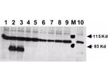
Rabbit anti-Human NOTCH 1 (Cleaved N Terminal) was used at a 1:500 dilution to detect mouse Notch 1 by Western blot. Equivalent amounts of lysates from transiently transfected 293 cells expressing recombinant myc-tagged mouse Notch constructs were electrophoresed and transferred to membrane using standard methods. A reaction with diluted primary antibody was followed by washing; reaction with a 1:10000 dilution of HRP conjugated Gt-a-Rabbit IgG (orb347654), and color development. Lane M: Mol wt markers. Lane 1: No transfection. Lane 2: N1 (mouse deleted extracellular domain)-myc. Lane 3: N1 (mouse intracellular domain)-myc. Lane 4: N2 (mouse deleted extracellular domain)-myc. Lane 5: N2 (mouse intracellular domain)-myc. Lane 6: N3 (mouse deleted extracellular domain)-myc. Lane 7: N3 (mouse intracellular domain)-myc. Lane 8: N4 (mouse deleted extracellular domain)-myc. Lane 9: N4 (mouse intracellular domain)-myc. Lane 10: N1 (mouse deleted extracellular domain)(V to G)-myc.

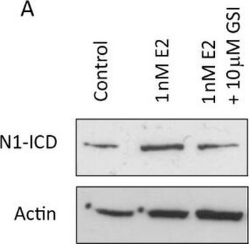
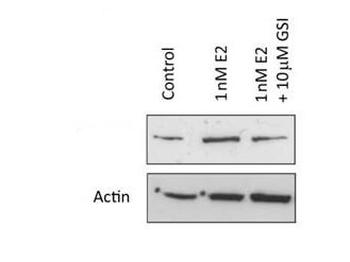
Systemic oestrogen signalling is mediated by EGFR and Notch. (A) Representative Western blot showing expression of cleaved (active) Notch1 (N1-ICD) following culture ± 1 nM 17β-estradiol ± 10 μM GSI. (Bi) Representative Western blot showing expression of Notch ligands in sorted MCF7 cells (left) and, where available, metastatic cells (right). (Bii) Densitometric analysis of three independent repeats of MCF7 sorting and of a single experiment for primary cells. Comparisons between population 1 (CSC enriched) and other populations are displayed. (C and D) Mammosphere formation was assessed following culture with 1 nM 17β-estradiol ± gamma secretase inhibitor (GSI) alone and in combination with gefitinib. Fold change is normalised to control, untreated cells represented as line. (E) Representative image of protein levels of ERK and phosphorylated (actived) ERK following culture for 48 hours in monolayer ± 10 μM GSI. Means plotted ± SEM, *P < 0.05, **P < 0.01, ***P < 0.001 compared to E2 treated. # P < 0.05 compared to control cells.

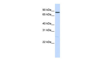
Western Blot of Rabbit anti-Notch1 antibody. Lane 1: MCF-7 control lysate. Lane 2: MCF-7 + 1 nM 17β-estradiol. Lane 3: MCF-7 + 10 μM gamma secretase inhibitor. Load: 35 µg per lane. Primary antibody: Notch1 antibody at 1:500 for overnight at 4°C. Secondary antibody: IRDye800™ rabbit secondary antibody at 1:10000 for 45 min at RT. Block: 5% BLOTTO overnight at 4°C. Predicted/Observed size: 80 kDa for Notch1.
- Item 1 of 7
NOTCH1 antibody [orb577050]
WB
Bovine, Canine, Equine, Guinea pig, Mouse, Rat, Zebrafish
Human
Rabbit
Polyclonal
Unconjugated
100 μl - Item 1 of 5
NOTCH1 antibody [orb750484]
DOT, ELISA, IF, IHC, IP, WB
Human, Mouse
Rabbit
Polyclonal
Unconjugated
200 μl - Item 1 of 4
- Item 1 of 4
- Item 1 of 4
Activated Notch1 antibody [orb312158]
FC, IHC-P, WB
Bovine, Canine, Guinea pig, Porcine, Rabbit, Rat
Human, Mouse
Rabbit
Polyclonal
Unconjugated
50 μl, 100 μl, 200 μl


Submit a review
Filter by Rating

- 5 stars

- 4 stars
- 3 stars
- 2 stars
- 1 stars
